
Abstract
Mobile phones create a radio-frequency electromagnetic field (EMF) around them when in use, the effects of which on brain physiology in humans are not well known. We studied the effects of a commercial mobile phone on regional cerebral blood flow (rCBF) in healthy humans using positron emission tomography (PET) imaging. Positron emission tomography data was acquired using a double-blind, counterbalanced study design with 12 male subjects performing a computer-controlled verbal working memory task (letter 1-back). Explorative and objective voxel-based statistical analysis revealed that a mobile phone in operation induces a local decrease in rCBF beneath the antenna in the inferior temporal cortex and an increase more distantly in the prefrontal cortex. Our results provide the first evidence, suggesting that the EMF emitted by a commercial mobile phone affects rCBF in humans. These results are consistent with the postulation that EMF induces changes in neuronal activity.
Introduction
Mobile phones in operation emit a pulsed radio-frequency electromagnetic field (EMF), a large part of the energy of which is absorbed into the user's head (Schonborn et al, 1998). Although the use of mobile phones is currently very common, there is scarce data concerning the effects of mobile phone use on brain physiology in vivo in humans. Some EEC studies have reported that EMF affects brain electrical activity (Reiser et al, 1995; Croft et al, 2002), especially during cognitive performance (Eulitz et al, 1998; Freude et al, 1998, 2000; Hamblin et al, 2004). In addition, some behavioural studies have suggested that EMF might have a facilitative effect on cognitive performance (Preece et al, 1999; Smythe and Costall, 2003; Koivisto et al, 2000a, b), although some recent studies have failed to show any effects of this kind (Haarala et al, 2003b, 2004). Our previous study using positron emission tomography (PET) yielded no conclusive evidence of any effect of the EMF emitted by a commercial mobile phone on regional cerebral blood flow (rCBF), although the subliminal noise emitted by the battery of the phone seemed to affect rCBF bilaterally in the auditory cortices (Haarala et al, 2003a). As our previous study aimed to examine interaction between EMF and cognitive task with a complex factorial design, including five different tasks, the statistical power concerning the main effect of EMF was not very strong.
The aim of the present study was to examine the main effect of EMF emitted by a commercial mobile phone on rCBF by using a substantially more sensitive experimental design than in our previous study (Haarala et al, 2003a). The confounding effects of noise from the battery of the mobile phone were eliminated by using a silent external power source. A double-blind, counterbalanced study with a within-subject design was conducted on 12 healthy humans performing a simple, computer-controlled working memory task during 14 PET scans, with the phone continuously in active EMF or sham mode during the first or second half of the scans. Working memory task was chosen as a ‘baseline', because a stable and easy task minimizes the random variation in rCBF between PET scans, and because task-related reaction times and error rates can be used to test the behavioural equivalence between exposure conditions. In addition, earlier experiments have reported effects of EMF during cognitive tasks in particular.
Materials and methods
Subjects
After written informed consent, 12 right-handed healthy male volunteers participated in the study, which had been approved by the local ethical committee. The average ± s.d. age, height, and weight of the subjects were 25 ± 2 years, 181 ± 8 cm, and 81 ± 10 kg, respectively. They abstained from caffeine, nicotine, and alcohol for 24 h before study and from mobile phone use on the day of the experiment. To exclude structural abnormalities of the brain, each individual had a 1.5 Tesla brain magnetic resonance imaging (MRI) (GE Signa, Milwaukee, WI, USA).
Electromagnetic Field Exposure
A factory model GSM (Global System for Mobile Communication) phone with the loudspeaker removed was used as the source of EMF exposure. In our previous experiments, we discovered a very low-level noise caused by the battery of the phone emitting EMF (see Haarala et al, 2003a). Although a measurement verified that this noise was below the human hearing threshold, our earlier results strongly indicated that even subliminal noise might induce a change in rCBF in the auditory cortices. Hamblin et al (2004), using the same mobile phone, also found a similar noise. Thus, to further eliminate any auditory effects, the battery was removed, and the phone was powered using a silent external power source positioned over 3 m away from the subjects. As the fully counterbalanced design eliminates all effects not related to the EMF exposure, an earplug was inserted into the subject's left ear to ensure that even immeasurably low noise from the phone in operation could not induce confounding auditory effects. The phone was placed between the subject's head and the head rest of the PET scanner parallel to the medial plane in such a way that the site of the removed loudspeaker was directly over the auditory canal, and the microphone was directed towards the corner of the mouth, that is, in the normal position of use (see Figure 1). The antenna was located approximately 17 mm from the surface of the skull over the subject's left inferior temporal lobe. A counterbalanced experiment with 10 independent subjects, each with 10 trials, indicated that the subjects could not detect the EMF exposure condition any better than by guessing (response accuracy 51%).
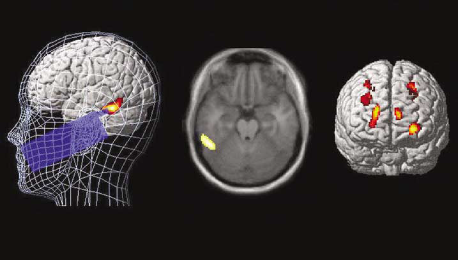
The position of the mobile phone and visualization of the results of a statistical analysis testing the effects of EMF exposure. (Left and centre panel) A cluster (pCorr value = 0.003; peak voxel's T = 4.94) with decreased rCBF during EMF exposure was located in the left fusiform gyrus in the posterior inferior temporal lobe (BA 37). (Right panel) Clusters with increased rCBF were located in the frontal lobe (pCorr values between 0.001 and 0.007, peak voxel's T-values between 4.53 and 5.52). See Table 1 for a detailed description of the results.
Results of the SPM analysis testing for differences in rCBF between the exposure conditions
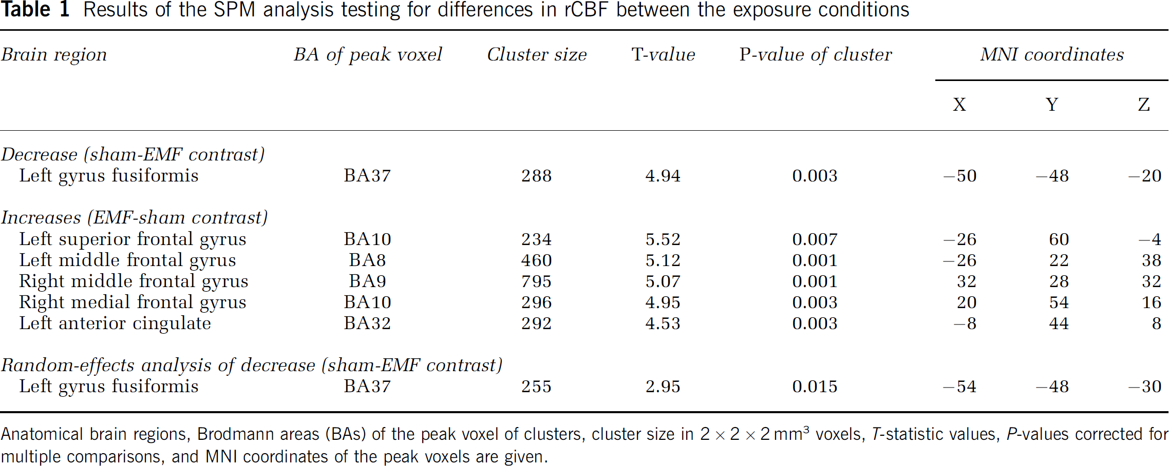
Anatomical brain regions, Brodmann areas (BAs) of the peak voxel of clusters, cluster size in 2 × 2 × 2 mm3 voxels, T-statistic values, P-values corrected for multiple comparisons, and MNI coordinates of the peak voxels are given.
The EMF emitted by the phone was controlled by mobile phone service software (WinTesla) with additional software to enable a double-blind design. When the EMF was on, the phone emitted a 902 MHz EMF with a mean power of 0.25 W pulsed at a frequency of 217 Hz with a pulse width of 0.577 ms. The specific absorption rate (SAR) of the phone was measured with the Dosimetric Assessment System 4 (DASY4; Schmid & Partner Engineering AG, Zürich, Switzerland) and a generic twin phantom filled with brain tissue-simulating liquid (σ = 0.969 Ω/m, εr = 40.14). SAR 10 g was 0.743 W/kg with an extrapolated peak value of 1.51 W/kg. The peak value was recorded near the position of the loudspeaker at the top part of the phone. Discontinuous transmission mode was not activated. The effect of the PET scanner on the intensity and distribution of SAR was investigated by using a SEMCAD V1.6 simulation platform and a high-resolution MRI base model. The simulation test revealed that the PET scanner increased the intensity of SAR 10 g by approximately 22%. The location of the peak SAR shifted by less than 1 mm.
Positron Emission Tomography Data Acquisition
A GE Advance (GE Medical Systems, Milwaukee, WI, USA) PET scanner in three-dimensional mode was used after a 200 MBq [15O]-water bolus injected into the left cubital vein. The acquisition of emission data was automatically started when the true coincidence rate exceeded 15 kcps. The EMF/sham exposure was started at the same moment as the transmission scan. After a 9-min transmission scan, each subject was scanned 14 times, out of which the first or last seven scans were obtained during a constant EMF or sham exposure in a double-blind, counterbalanced order. During both the former and the latter exposure conditions, the interval between the scans was 6 mins and that between the exposure conditions 15 mins (for timing issues, see Chmielowska et al, 1999). Thus, both the first and the last exposure periods were 51 mins. Each PET scan was integrated into a single 60-secs frame. The images were reconstructed with a filtered back-projection algorithm.
Behavioural Task
During both exposure conditions, the subjects performed continuously a simple working memory task (1-back task) controlled by the CogniSpeed program (Version 2; AboaTech Ltd, Turku, Finland). In this task, the subject is shown letters, one at a time, on a computer screen. They are instructed to respond by pressing a designated ‘Yes' key when the letter shown is the same as the previous letter, and by pressing a ‘No’ key in other instances. The same stimulus set (sequence of letters) was used in both exposure conditions, to ensure that there was no cognitive difference between them.
Statistical Image Analysis
The preprocessing and statistical image analyses were performed using the Statistical Parametric Mapping (Friston et al, 1995) software version 99 (SPM99) and Matlab 6.5 for Windows (Math Works, Natick, MA, USA) with a previously described procedure (Aalto et al, 2002). Briefly, after realignment (motion correction) of each subject's image series, the normalization parameters for each subject were estimated from the mean image using a default algorithm and a [15O]-water PET template delivered with SPM99. To improve the signal-to-noise ratio and to reduce the residual interindividual neuroanatomic variation, the images were smoothed with an isotropic Gaussian filter of 12 mm full-width at half-maximum. Subtraction analysis testing the effects of EMF exposure (EMF versus sham) was performed using a multisubject condition and a covariate model. The effects of the variation in global CBF (gCBF) were eliminated by subject-specific global normalization of the analysis of covariance (by subject), which labels gCBF as a nuisance covariate. Applying the default settings, the mean voxel values were subject specifically scaled to 50 mL/dL per minute, and a proportional analysis (gray matter) threshold of 0.8 was applied. The model specifications resulted in a design with 26 parameters, leaving 143 degrees of freedom. Statistical testing was performed using exploratory fixed-effect analysis covering the entire brain without any a priori hypothesis about the location of effects. The search volume consisted of over 200,000 voxels forming 508 resolution elements (resels). An additional SPM analysis utilizing the random-effects model was performed to evaluate whether it is possible to find near the peak SAR area an effect generalizable to the population level. The detailed analysis procedure has been described earlier (Aalto et al, 2005b). Briefly, the two-phase analysis procedure included a t-test of contrast images generated by subject-specific subtraction analysis at the first phase of analysis. To enhance the sensitivity of the analysis, the search volume was constrained to the circular area with about 3 cm radius surrounding the cortical surface beneath the base of the antenna. The search volume was defined on the MRI template image using the Imadeus software 1.50 (Forima Inc., Turku, Finland) with a standard procedure (e.g., Aalto et al, 2005a, b ) and included 3954 voxels (31.6 cm3). P-values, corrected for multiple comparisons, lower than 0.05 were considered statistically significant. For localization, the coordinates of the maxima of the clusters resulting from statistical analysis were converted into Talairach coordinates (Talairach and Tournoux, 1988) using the mni2tal conversion software by Brett (http://www.mrccbu.cam.ac.uk/Imaging/mnispace.html) and localized using the Talairach Deamon Software (Lancaster et al, 2000).
Results
The statistical analysis testing the effects of EMF exposure revealed a decrease in rCBF during the ‘EMF’ state in the left fusiform gyrus in the posterior inferior temporal cortex below the antenna (Figure 1, left and centre panel), while an increase in rCBF was seen mainly bilaterally in the superior and medial frontal gyri (Figure 1, right panel). The additional random-effects analysis restricted to the area covering roughly the most intensive SAR area revealed a cluster with decreased rCBF in the left fusiform gyrus. A detailed description and anatomic localization of the results of the statistical analyses are presented in Table 1. The EMF had no effects on reaction times (T11 = 0.602, P = 0.559) or the accuracy of responses (T11 = 0.936, P = 0.369). Also, the reaction times and the accuracy of responses behaved fairly similarly as a function of time during both conditions (Figure 2).
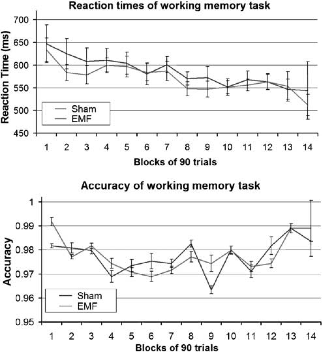
Mean reaction times and accuracy of the working memory task during exposure conditions. The mean and s.e.m. values were calculated in 90-trial blocks during continuous repetition of a 1-back working memory task. There are only minor and random differences in the reaction times and the accuracy of the responses between the conditions. The temporal trends of these variables also seem to be fairly similar during the conditions.
Discussion
Although, in the field of cognitive neuroscience, it is generally assumed that changes in rCBF reflect changes in neuronal activity (see, e.g., Jueptner and Weiller, 1995), this interpretation is not self-evident in the presence of EMF exposure. Indeed, it is possible that the observed changes are caused by some vascular effects of EMF. However, even near the antenna, thermal mechanisms of a decrease in local rCBF are unlikely, as the rise in brain tissue temperature immediately below an active mobile phone antenna is only 0.11°C, and a rise in temperature should increase rCBF (Van Leeuwen et al, 1999). Thus, neuronal mechanisms seem more likely. In vivo animal and in vitro experiments have suggested some possible mechanisms for EMF effects on neurons, such as changes in cell membrane permeability, calcium efflux, or neuronal excitability, but controversies remain (for a thorough review, see Independent Expert Group on Mobile Phones, 2000).
As the EMF emitted by a mobile phone attenuates very quickly, especially in tissue (Schonborn et al, 1998), it is not probable that the effects found in the prefrontal cortex were caused by any direct effects of EMF. Also, the fact that the frontal effects consisted of increases in rCBF, unlike the local effect, which was a decrease, suggests different mechanisms behind these ‘remote’ effects. Baseline task-related EMF effects are not probable, as these effects were not located in the regions typically found to be involved in verbal working memory, namely the Brodmann areas (BA) 6, 44, and 46 (for a review, see Cabeza and Nyberg, 2000). Thus, it is more probable that the frontal increases in rCBF reflect increased neuronal activity mediated by neural connections between the frontal areas and the left inferior temporal areas near the peak EMF (see, e.g., Bitan et al, 2005; Gazzaley et al, 2004; Powell et al, 2004)
We suggest that the rCBF changes found in this study are related to EMF-induced changes in neuronal activity. However, these changes do not seem to have influenced the task at hand, as the EMF had no significant effects on reaction times or the accuracy of responses. Thus, our results do not support the assumption that changes in neuronal activity would be related to facilitation in cognitive performance induced by EMF exposure, which has been suggested in some previous studies (Preece et al, 1999; Smythe and Costall, 2003; Koivisto et al, 2000a, b). Our negative behavioural results as such are in line with the most recent studies failing to replicate the earlier findings of cognitive facilitative effects of EMF exposure (Haarala et al, 2003b, 2004).
In addition to our previous study (Haarala et al, 2003a), there is only one PET study examining the effects of EMF on rCBF in humans (Huber et al, 2002, 2005). However, any comparison of the results of that study and the present study is difficult, as the signal generator with a planar antenna used by Huber et al creates EMF exposure with different characteristics than the commercial mobile phone used in our study. More importantly, Huber et al acquired all of their PET scans after the EMF exposure, whereas we did the scanning during the exposure, as the immediate effects of the EMF were considered more probable based on previous studies. However, Huber et al (2002) reported markedly increased rCBF in the ipsilateral prefrontal cortex and the contralateral parietal cortex without any effect near the peak SAR area. The unbalanced study design and the uncontrolled baseline task (silent counting) make the interpretation of these results very difficult.
There are not many probable confounding factors that could have affected the results of the present study. The design was double-blind and fully counterbalanced, eliminating all periodic effects as well as the effects of the baseline task as such. In line with this, our findings are not functionally explainable by the task at hand. On the contrary, decreased rCBF near the antenna was located in the gyrus fusiformis, which is activated by the letter recognition called for by the baseline task (e.g., Polk et al, 2002), However, the behavioural results indicated that neither the EMF nor other factors caused such changes in cognitive performance that would plausibly explain our findings. After all, as the statistical analysis was made using a within-subject crossover design, the findings cannot be based on sampling differences. However, one should note that, when the fixed-effect model is used, effects at the population level cannot be estimated. Although, the confirmatory random-effects analysis indicated that the local decrease in rCBF in the high SAR area in the fusiform gyrus can be reliably generalized to the population level, we emphasize that replication studies with a larger sample are warranted.
Conclusions
To our knowledge, this is the first brain imaging study suggesting that EMF, per se, emitted by a commercial mobile phone induces changes in rCBF. Although the cellular mechanisms behind these findings are unclear, these results are consistent with the interpretation that EMF induces changes in neuronal activity. However, we want to underline that our results do not provide any evidence to suggest that the use of mobile phones would be more harmful to brain tissue than normal cognition, which is also always accompanied by intense temporary changes in neural activity and rCBF.
Footnotes
Acknowledgements
We thank professor Matti Laine (Åbo Akademi University, Turku, Finland) and Liisa Metsähonkala, MD, PhD, (Turku University Hospital, Turku Finland) for valuable comments on the manuscript and the staff of the Turku PET Centre for assistance in imaging.