
Abstract
Functional magnetic resonance imaging (fMRI) using paced auditory serial addition test (PASAT) as paradigm was used to study the functional connectivity in 18 patients at the very early stage of multiple sclerosis (MS) compared with 18 controls, to determine the existence of circuitry disturbance inside the working memory network and its relationship with white matter abnormalities assessed by conventional MRI and magnetization transfer ratio (MTR) imaging. The left BA 45/46 was selected as the seed region to compute correlation maps with other brain regions. After obtaining the correlation map for each subject, between-group comparisons were performed using random effect procedure. Compared with controls, patients did not show any greater functional connectivity between left BA 45/46 and other regions during PASAT. In contrast, decrease in functional connectivity was observed in patients between left BA 45/46 and left BA 9, right BA 3, and the anterior cingulate cortex (BA 24). In patients, no correlations were found between altered functional connectivity and clinical data. However, functional connectivity observed between left BA 45/46 and BA 24 in patients was correlated with the MTR of normal appearing white matter, and with brain T2 lesion load. Altered functional connectivity is present inside the working memory network of patients at the very early stage of MS and is related to the extent of diffuse white matter changes.
Introduction
Neuropsychological studies have shown that cognitive impairment is a common finding in multiple sclerosis (MS), detected in 40% up to 60% of patients (Heaton et al, 1985; Lyon-Caen et al, 1986; Peyser et al, 1980; Rao et al, 1991). In patients with long lasting disease duration, brain atrophy appears partly related to the extent of cognitive impairment. At this stage, axonal loss may constitute an important factor contributing to this impairment. But at the early stage of the disease, namely in patients with clinically isolated syndrome (CIS) suggestive of MS, diffuse myelin destruction could also play an important role, when several magnetic resonance spectroscopy (MRS) studies have reported absence of neuronal loss markers (Fernando et al, 2004; Ranjeva et al, 2003). At the very early stage of MS, cognitive impairment appears limited to attentional, working memory and conflict monitoring capacity (Achiron and Barak, 2003; Pelosi et al, 1997), and is highly related to the decrease of magnetization transfer ratio (MTR) inside the normal appearing white matter (NAWM) (Audoin et al, 2004; Ranjeva et al, 2005). Up to now, the neuropathologic substrate of cognitive impairment at the early stage of MS has remained largely unknown according to the fact that no histopathologic data are available. One assumption is that disruption of neural connections among cortical associative areas occurring as a consequence of diffuse white matter damage, may constitute the anatomical substrate of cognitive impairment in early MS. Supporting this assumption, long distance cortico-cortical connections appear particularly involved in various high cognitive processes like attention, working memory, and consciousness. The ‘conscious neuronal workspace’ theory proposed by Dehaene et al (2003) emphasizes the role of distributed neurons with long-distance connections, particularly dense in prefrontal, cingulate, and parietal cortices, that is, regions implied in executive processes that are capable of interconnecting multiple specialized processors during conscious processes. In this context, the integrity of WM fiber tracts could be a limiting factor to perform correctly high controlled information processing tasks involving several distant brain areas that have to interact synchronously.
For more than a decade, functional magnetic resonance imaging (fMRI) has brought new insight into the better understanding of brain functioning by its ability to observe noninvasively activation patterns during a given task. In addition, information relative to the organization of the cerebral networks can also be assessed by computing effective connectivity and/or functional connectivity from the same fMRI data set. While most of the studies using these fMRI-derived parameters have been conducted separately, information brought by each analysis (activation pattern, functional connectivity, and causal interactions) appear to be complementary and nonredundant. In this context, determining on the same set of fMRI data different functional parameters could lead to the better characterization of brain functioning in normal but also in pathologic states.
We have recently reported in a population of patients at the very early stage of MS, differences in fMRI activation patterns during a working memory task (PASAT) compared with controls (Audoin et al, 2003, 2004). These functional changes were located in regions involved in executive processing (prefrontal cortices). Activation in the right lateral prefrontal cortex (LPC) has also been inversely correlated with the extent of diffuse tissue damage evaluated by MTR imaging (Audoin et al, 2004). Structural equation modeling (s.e.m.) applied on the same fMRI data set has also allowed to show differences in causal interactions between cortical areas assumed to take part in the working memory network (effective connectivity) (Au Duong et al, 2005). Modulation of effective connectivity in CIS patients was present with both decrease and increase in causal interactions inside the executive systems of working memory (Au Duong et al, 2005), where comparisons of activation maps evidenced only diffuse activation enhancement (Audoin et al, 2003, 2004). However, differences in effective connectivity between patients and controls could only be observed along the predefined links included in the arbitrary cognitive model of working memory, whereas other networks or connections potentially implied in the task would have been skipped.
In contrast, functional connectivity has the advantage of not precluding any possible connectivity organization of a given cortical network. Indeed, functional connectivity is defined as the temporal correlation between different neurons in a given system (Friston et al, 1993). It could reveal direct connection between two areas as well as indirect connection mediated by areas not known. Several studies have reported the use of fMRI data to assess functional connectivity in healthy subjects during resting conditions (Xiong et al, 1999; Greicius et al, 2003; Jiang et al, 2004), or during a specific task involving the motor system (He et al, 2003; Rogers et al, 2004), language organization (Hampson et al, 2002; Homae et al, 2003), and the visual and auditory systems (Horwitz and Braun, 2004). Concurrently, differences in functional connectivity have been showed in patients with schizophrenia compared with controls (Lawrie et al, 2002). This approach has also been used in clinically definite MS patients investigating the motor cortex (Lowe et al, 2002; Saini et al, 2004). However, no functional connectivity data using fMRI acquisitions are currently available concerning the high-level cognitive systems in patients with MS. To evaluate the impact of early MS on the integrity of the neural networks involved during PASAT, functional connectivity has been determined in the same fMRI data set recorded in 18 CIS patients and 18 controls.
Materials and methods
Subjects
We have used functional and structural MRI data from 18 patients with CIS fulfilling at inclusion at least the dissemination in space criteria of MS according to McDonald (dissemination in space showed by MRI, or positive cerebrospinal fluid plus two or more MRI detected lesions consistent with MS). At the time of inclusion in the study, 14 patients had a diagnosis of MS and four had a diagnosis of ‘possible MS’ according to McDonald's criteria, when comparing the first MRI performed after relapse and conventional MRI performed at the same time as fMRI exploration (at least 3 months after onset and steroid treatment). Follow-up scans using the same conventional MRI procedures, performed 6 months and 1 year later, showed conversion of the four ‘possible MS’ patients into ‘MS’ according to the time evolution of the disease (new T2 lesions and/or gadolinium-enhanced lesions). The functional status of subjects was evaluated using the expanded disability status scale (EDSS) (Kurtzke, 1983) and the multiple sclerosis functional composite (MSFC) score (Cutter et al, 1999; Fischer et al, 1999). The 18 patients were matched for age, sex, and educational level with 18 healthy controls. The control group was used as reference for the calculation of the MSFC score. All subjects (patients and controls) were right-handed (>70% Oldfield scale) (Oldfield, 1971), native French speakers and naive people with respect to PASAT. They gave their informed consent for their participation in this experiment that was approved by the local Ethics Committee (Timone Hospital, Marseille, France).
Conventional and Structural Magnetic Resonance Imaging
Magnetic resonance imaging acquisition: Images were acquired using a 1.5 T Magnetom Vision Plus MR imager (Siemens, Erlangen, Germany). The MRI protocol included localizer scout images, transverse fast SE proton density-weighted and T2-weighted images (TE1/TE2/TR = 15/85/2600 ms; 44 contiguous slices, thickness = 3 mm; flip angle = 90°; FOV = 240 mm; matrix = 2562), transverse proton density-weighted spoiled gradient echo sequences (TE/TR = 4.7/500 ms, 44 contiguous slices, thickness = 3 mm, flip angle = 30°; FOV = 240 mm; matrix = 2562) performed without and with MT saturation (1.5 kHz off water resonance, flip angle = 500°). A T1-weighted SE sequence (TE/TR = 10/650 ms, 25 contiguous slices, thickness = 5 mm; flip angle 90°, FOV = 240 mm, matrix = 2562) was also performed before and 5 mins after injection of gadolinium (0.1 mmol/L kg).
Magnetization transfer ratio image processing: Magnetization transfer ratio maps were calculated on a voxel-by-voxel basis according to the following equation: MTR(%) = (M0–Mmt)/M0, where M0 and Mmt were the images obtained without and with MT saturation pulse, respectively. Magnetization transfer ratio maps were coregistered onto the corresponding T2-weighted images. Means MTR of the NAWM were obtained from histograms described in previous works (Audoin et al, 2004; Ranjeva et al, 2005). Comparison of mean NAWM MTR between patients at the very early stage of MS and controls was assessed using nonparametric Mann—Whitney U-Test.
Functional Magnetic Resonance Imaging
Design: PASAT task consisted in auditory entry series of 61 single-digit numbers delivered every 3 secs (PASAT 3). After delivery of each number (except the first), subjects were asked to overt vocalize the result of the addition of the two last numbers heard. Performances of subjects were recorded during fMRI by an investigator present in the examination room and were used to compute the MSFC score of each subject. This fMRI paradigm using a long sustained period of activation (60 consecutive measurements) is well suited to determine the temporal signal correlations between several cortical areas for functional connectivity analysis, avoiding the signal gaps induced by task alternances. Furthermore, sustained concentration is a major component of conventional PASAT, making this task difficult to realize.
Data acquisition: Images were acquired using the same scanner as for structural MRI using single-shot gradient-echo EPI sequence (TE = 54 ms, bandwidth 1470 Hz/pixel, 17 slices, 5 mm thickness, 0.5 mm distant factor, matrix = 1282, FOV = 240 mm, pixel size = 1.882 mm2). Acquisition time for a block of 17 slices was 2.25 s. EPI blocks were acquired at a rate of one block every 3 secs. The 61 single-digit numbers (stimulus duration = 500 ms) were delivered using a personal computer running the Cool Edit Pro software (version 1.2, Syntrillium software corporation, Phoenix, AZ, USA), every 3 secs during the intervals of silence (blank duration = 750 ms). A morphological 3D-T1 weighted magnetization prepared rapid gradient echo (MPRAGE) (TE = 4.7 ms, TR = 9.7 ms, flip angle = 12°, 128 partitions, matrix = 2562, isotropic voxel 1.253 mm3) was also acquired to superimpose statistical maps.
Image preprocessing: Images were processed using the SPM99 software (Wellcome Institute, London, UK). After realignement, images were normalized, coregistered and 8 mm-smoothed with a Gaussian filter.
Data Analysis
Data from published functional imaging studies (Smith and Jonides, 1999), and from our previous fMRI studies using PASAT as paradigm (Audoin et al, 2003, 2004) emphasized the key role of the left LPC (BA 45/46) in the verbal working memory system, especially in the executive system. Thus, we focused on the functional connectivity involving this area. Using the marsbar toolbox (SPM 99), we defined the source ROI by selecting the intersection of the clusters obtained in the two groups of patients and controls and belonging to the left BA 45/46 that were significantly activated during PASAT both in patients and controls (P<0.005, spatial extent k>10).
For each subject, we averaged the time-series of the voxels included in the source ROI and we extracted, from this average, the signal corresponding to the PASAT period. The resulting time-series was used as covariate of interest in a linear regression model with time-series of each brain pixel during PASAT. A correlation map (r-map) for each subject was then obtained.
Subject-specific r-maps were then entered into second-level within-group analysis (one sample t-test P<0.0005 corrected, k>10) to obtain the functional connectivity network of each group, and between-group analysis (two sample t-test P<0.001, k>10) to determine significant differences between patients and controls in functional connectivity during PASAT.
Individual r-maps of patients were also used to study correlation between functional connectivity and brain T2 lesion load, mean MTR NAWM, PASAT performance, MSFC and disease duration.
Results
Clinical Characteristics
Comparisons of group characteristics are summarized in Table 1. Patients and controls showed no statistical differences related to age, sex, and educational level. Multiple sclerosis functional composite was significantly reduced in patients (P = 0.04), according to a decrease in PASAT scores (P = 0.004), with nonsignificant differences in the nine-hole peg test (P = 0.323) and in the 25-feet walk (P = 0.0905). A low median EDSS of 1 (range 0 to 2) was observed in patients with a moderate mean T2 lesion load of 2.15±1.42 cm3.
Comparative demographics and clinical characteristics of patients and controls
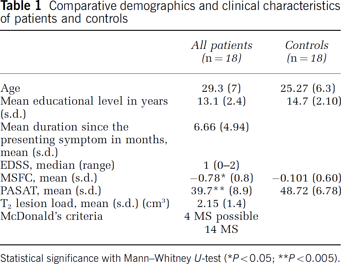
Statistical significance with Mann—Whitney U-test (*P<0.05; **P<0.005).
Characteristics of Tissue
Statistical comparisons (Mann—Whitney U-test) showed that mean NAWM MTR values were significantly lower in patients (46.81%±1.00%) compared with controls (47.59%±0.53%) (P<0.05). In addition, in patients, mean NAWM MTR values were significantly correlated with the PASAT scores (r = 0.658, P = 0.0068).
Left Lateral Prefrontal Cortex Functional Connectivity
Brain areas with significant functional connection with left BA 45/46 in healthy controls are summarized in Table 2. The most connected areas are in accordance with previous fMRI studies of working memory (Smith and Jonides, 1999). Quantitatively, patients show significant lower functional connectivity between the left BA 45/46 and the left BA 9, the right BA 3 and the anterior cingulate cortex (ACC) (BA 24) compared with controls (Figure 1). In contrast, patients do not show any greater functional connectivity between the left BA 45/46 and other brain areas compared with controls.
Brain areas displaying significant functional connectivity with left BA 45/46 in 18 healthy controls (random effect analysis, one sample t-test P<0.0005 corrected, k>10)
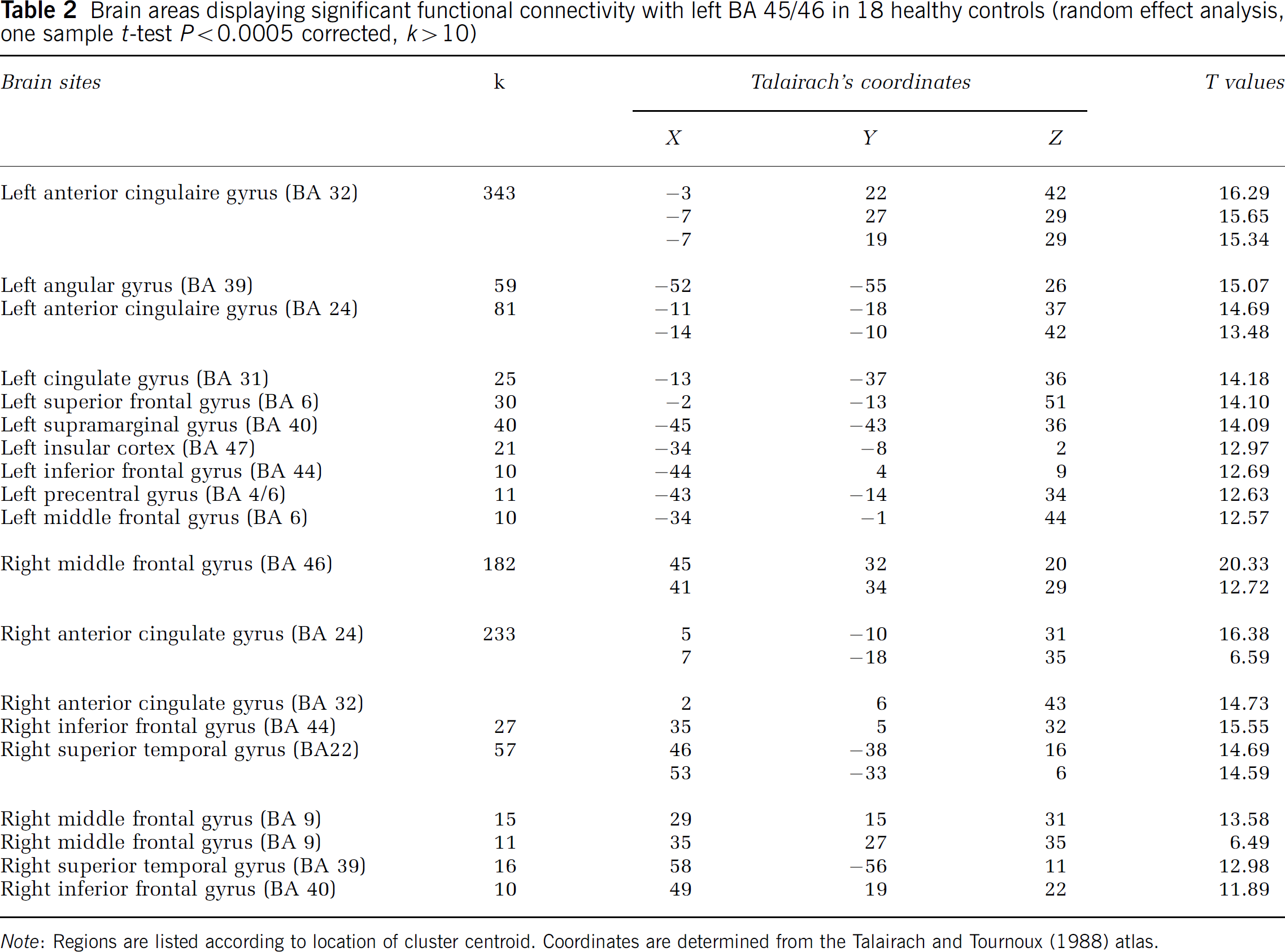
Note: Regions are listed according to location of cluster centroid. Coordinates are determined from the Talairach and Tournoux (1988) atlas.
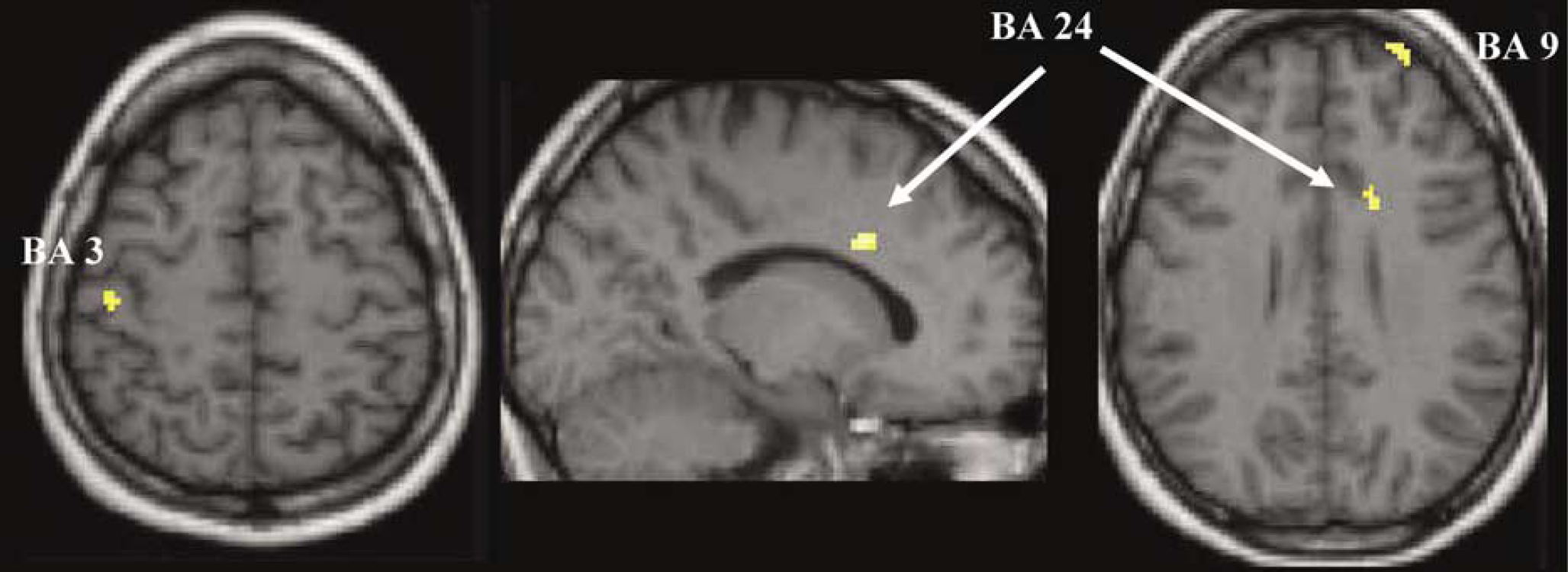
Location of significant decreases in functional connectivity during PASAT observed in patients with early multiple sclerosis (MS) compared with controls (left prefrontal cortex as seed region). After computing correlation maps (r-maps) during PASAT for each subject with the temporal functional magnetic resonance imaging (fMRI) signal from the left BA 45/46 as covariate, between-group comparison of individual correlation maps (random effect, two sample t-test P<0.001, k>10) showed a relative decrease in functional connectivity affecting links between the left BA 45/46 and the left BA 9, the left BA 45/46 and the right BA 3, the left BA 45/46 and the anterior cingulate cortex (ACC) (BA 24).
In patients, no correlations were found between altered functional connectivity and clinical data (PASAT, MSFC, disease duration). In contrast, significant correlations were found between altered functional connectivity linking the left BA 45/46 with the ACC, on the one hand the mean MTR NAWM (P<0.005, k>10) (Figure 2), and on the other hand the brain T2 lesion load (SPM analysis using linear regression P<0.005, k>10); confirmed with Spearman's rank test on extracted data (P<0.05) (Figure 3).
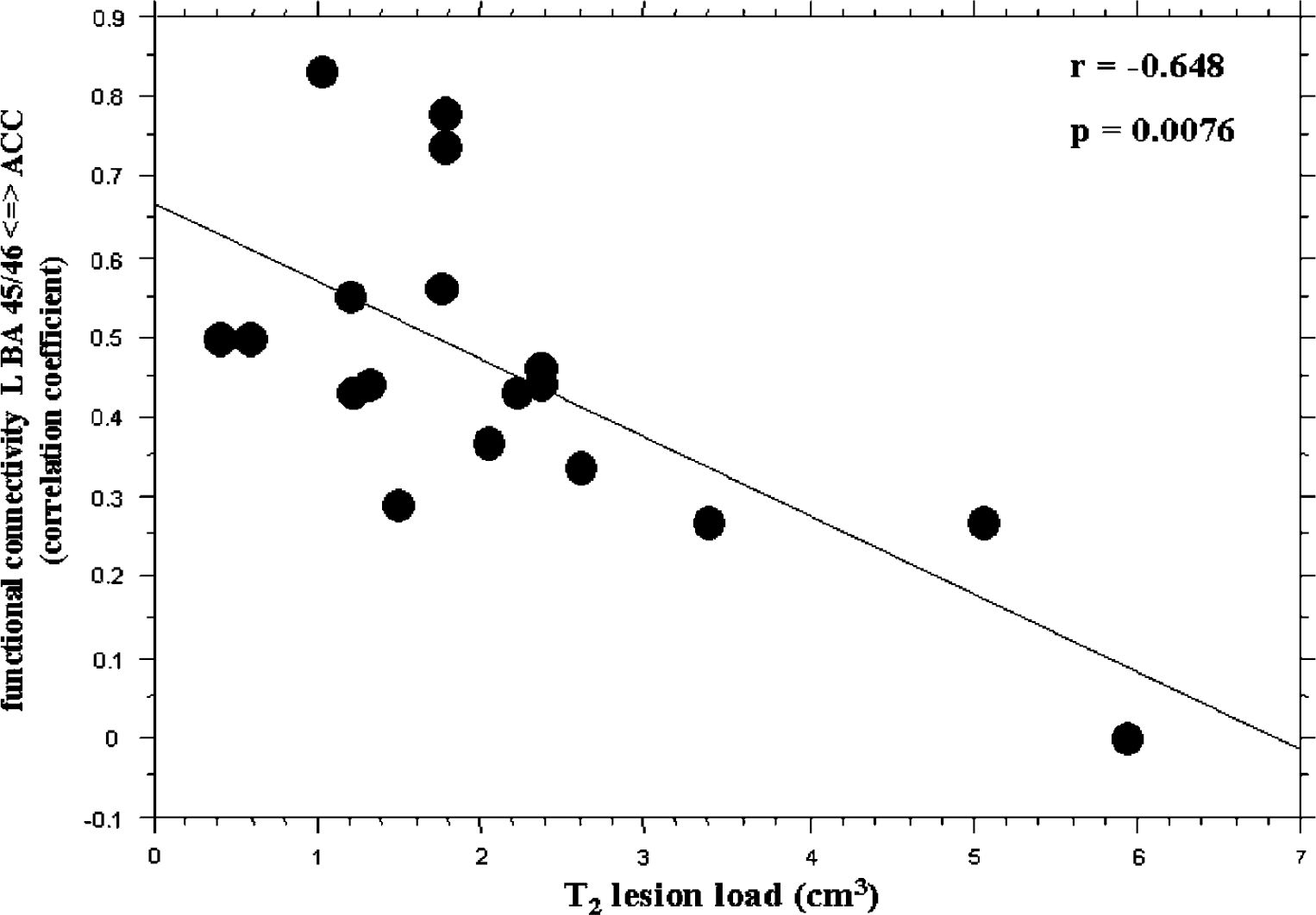
Correlation between the T2 lesion load and the functional connectivity (correlattion coefficients) evaluated between the left BA 45/46 and the anterior cingulate cortex (Spearman's rank test).
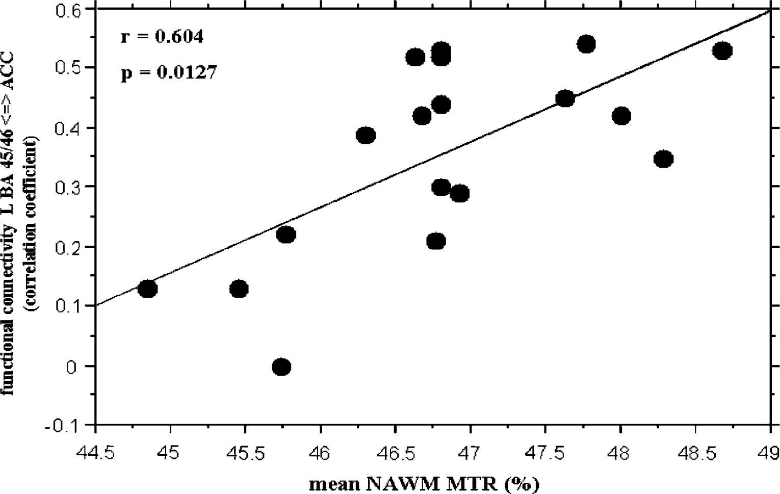
Correlation between the mean normal appearing white matter (NAWM) magnetization transfer ratio (MTR) values and the functional connectivity (correlation coefficients) evaluated between the left BA 45/46 and the anterior cingulate cortex (Spearman's rank test).
Discussion
Technical Aspects and Limitations
Experimentally, functional connectivity is evaluated from temporal correlations between one measured signal supposed to reflect to some extent the neuronal activity during a given task inside one seed region (left BA 45/46), and the corresponding signals measured during the same period in the other parts of the brain. Some technical issues should be considered in the interpretation of these data, since fMRI does not directly measure synaptic changes, but indirect relative variations in blood flow. Temporal correlations obtained from fMRI acquisitions can potentially also be related to other mechanisms independent from real functional connectivity such as head motions. As long as subjects were asked to vocalize responses, we cannot totally exclude the presence of signal changes because of head motions, inducing voxel displacements or variations in local magnetic susceptibility. Some authors have proposed to handle those issues by using independent component analysis (ICA) techniques to remove unexpected components (McKeown et al, 1998a, b ). However in the present study, the overt responses were composed by one or maximum two syllabi (simple numbers from 2 to 18). Vocalization of these simple words did not induce large head motions and transformations provided by realignment procedures did not exceed 5 mm in each direction for each subject. Moreover, variations in susceptibility artefacts that could lead to false-positive differences between groups would have preferentially occurred in the temporal or occipital lobe rather than in the anterior frontal lobe where the significant differences were actually observed. It has also to be noted that the long sustained period of activation used in this paradigm (60 consecutive measurements) has appeared well-suited to accurately determine the correlations of temporal signals between several cortical areas, avoiding signal gaps because of concatenation of fMRI signals recorded during several distant periods because of alternance of conditions. As the regions shown to have lower functional connectivity with the left BA 45/46 in patients (the left BA 9, the right BA 3 and the ACC) were not homologous and not adjacent, these correlations could not be attributed to the symmetry of the diminution in the blood supply of these regions or to the low spatial resolution of functional MRI. It could be also noted that these regions were not the most sensitive to signal changes related to magnetic susceptibility variations induced by motion, giving an additional confidence onto the results. The random effect analysis has finally helped to minimize the influence of inter-subject variability and has also given the opportunity to obtain individual parameters used to determine correlations between altered functional connectivity and structural or clinical parameters.
Brain Functioning During PASAT in Patients at the Very Early Stage of Multiple Sclerosis
Performing PASAT mobilizes several cognitive systems and appears very demanding on verbal working memory, because subjects have to (i) maintain verbal information (the two last numbers heard) during a short period of time, and (ii) manipulate this information by adding these two numbers (Audoin et al, 2003). In agreement with the model proposed by Baddeley (1986, 1992, 2003), numerous imaging studies in healthy subjects have identified cortical areas particularly implied in the working memory network (Cabeza et al, 1997; Della-Maggiore et al, 2000; Krause et al, 2000; Table 1Glabus et al, 2003; Schlosser et al, 2003; Kondo et al, 2004). There is evidence that the ACC and the left LPC (BA 46) are predominantly subserving executive control functions of verbal working memory, whereas preprocessing and maintenance of verbal information are mainly associated with activation of the left ventrolateral frontal (BA 44) and also the left parietal associative cortex (BA 40) (Collette and Van der Linden, 2002; D'Esposito et al, 1995; Duncan and Owen, 2000; Osaka et al, 2003; Paulesu et al, 1993; Smith and Jonides, 1999). It has been emphasized that the ACC and LPC are important components of the distributed attentional network (Posner, 1994; Posner and Dehaene, 1994). In particular, ACC could particularly be involved in conflict monitoring and cognitive control whereas the dorsolateral prefrontal cortex is involved in top down processes. Closer cooperation between the two brain regions appears strongly related to working memory performance (Kondo et al, 2004).
In the current study, functional connectivity measured in controls during PASAT using the left BA 45/46 as seed region, has shown links between the LPC and most of the regions reported to play a role in the working memory network (Smith and Jonides, 1999). In patients at the very early stage of MS, altered functional connectivity has been observed inside the working memory network, with a decrease in functional connectivity affecting connections linking the left lateral prefrontal cortex to the ACC, to the left BA 9 and to the right BA 3. Impaired functional connectivity between the left LPC and the ACC observed in the present study was in accordance with the decrease in causal interactions from the ACC to the left BA 45/46 previously observed in the same population (Au Duong et al, 2005).
Moreover, the extent of diffuse white matter damage expressed by T2 lesion load and NAWM MTR has been correlated to the functional connectivity between the left lateral prefrontal cortex and the left ACC. This correlation could potentially be explained by conduction disturbances, related at least in part to myelin damage, and/or disruption of WM fibers (axonal damage) between these two cortical regions. Decrease in functional connectivity could then be considered as a surrogate marker of structural integrity of the brain network. These ‘structural dimensions’ of functional connectivity could also explain the absence of increased functional connectivity in patients compared with controls, while increased causal interactions were observed using effective connectivity analysis on the same population (Au Duong et al, 2005). However, striking is the fact that no direct correlations have been observed between the altered functional connectivity and PASAT scores, showing that disturbances between cortical areas involved in executive processes fail to explain alone the cognitive impairments. This lack of correlation was also observed between the T2 lesion load and PASAT scores, while NAWM MTR was significantly correlated with PASAT, explaining approximately 45% of the variance. One possible explanation could be related to mechanisms involving network reorganization inside the working memory system and/or compensatory cortical activation known to occur at this stage of the disease. Increase in activation inside the prefrontal cortices (Audoin et al, 2004, 2003) and modulation of causal interactions between several cortical areas of executive systems of working memory (Au Duong et al, 2005) previously observed, could be related to compensatory mechanisms that could tend to limit the impact of injured brain circuitry.
In conclusion, altered functional connectivity is present inside the working memory network of patients at the very early stage of MS and is related to the extent of white matter tissue damage. Further study using multiparametric modeling combining different aspects of tissue and functional pathologic brain substrate would help to better understand the cognitive impact of diffuse white matter damage. More studies will also be necessary to document the pathologic substrate of white matter MTR decrease in early MS.