
Introduction
Cerebral vascular mean transit time (MTT), calculated as the ratio of cerebral blood volume (CBV) to cerebral blood flow (CBF), is expected to be inversely proportional to cerebral perfusion pressure. Dynamic susceptibility contrast-enhanced magnetic resonance imaging (DSC-MRI) with a Gd-based contrast agent is being applied increasingly for cerebral perfusion study, although positron emission tomography (PET) measurement has been regarded as the gold standard for quantification of CBF. The objective of this study was to examine the difference between MTT determined by PET (PET-MTT) and that determined by DSC-MRI (MRI-MTT).
Materials and Methods
Subjects were seven healthy volunteers, 20–21 years of age. In the PET study, the CBV image was derived from scanning after [15-O]-CO inhalation. CBF was calculated by least-squares fitting of the dynamic data obtained with [15-O]-H2O bolus injection and continuous arterial sampling. In the DSC-MRI study with bolus injection of Gd-based contrast agent, dynamic data were obtained with a 1.5 T scanner at 1-second intervals using a gradient-echo EPI. CBV was calculated by integrating the time-concentration curve. CBF was determined with the use of singular value decomposition deconvolution 1 incorporated with correction for the effect of tracer delay 2 . Regions of interest 10 mm in diameter were drawn on the PET and DSC-MRI images.
Results and Discussion
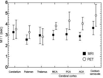
Average MTTs are shown in Figure 1. The PET data were consistent with previously reported PET data in terms of regional distribution of MTT 3 . The MRI-MTT differed regionally, as did the PET-MTT. However, MRI-MTTs were systematically shorter than PET-MTTs except in the thalamus. A possible explanation for this difference is the sensitivity of each imaging modality to vascular components: PET measurements for CBV are obtained with the use of [15-O]-CO and are equally sensitive to all vascular components, whereas DSC-MRI signals originate from the microvasculature in vicinity to the brain parenchyma. Thus, MTT obtained via DSC-MRI should be shorter than MTT obtained via PET imaging. Therefore, the two sets of data appeared consistent, although potential sources of error in the two methods should be carefully considered.
Footnotes
Acknowledgements
This research was supported in part by a Ministry of Education, Science, Sports and Culture Grant-in-Aid for Young Scientists (16790751).