Introduction
Basis function methods (BFM) have been developed for generating images of binding potential and perfusion1, 2, 3. These methods are based on a one-dimensional (1D) series of basis functions. In the present study a 2D BFM was developed for generating binding potential (BP) and volume of distribution (Vd) images based on a two tissue compartment plasma input model.
Methods
The time activity curve of a two tissue compartment model can be described by convolution of the plasma input with 2 exponentials. The 2D BFM is based on pre-computing both convolutions for a series of discrete exponential values, i.e. 2 sets of basis functions are generated. For each combination of basis functions, the optimal set of fit parameters and the corresponding value of the cost function can be obtained by simple linear regression. The combination of basis functions and fit parameters, which provides the lowest value of the cost function, can then be used to calculate BP and Vd. A simulation study was performed to study the effects of noise on accuracy and precision of BP and Vd obtained with this 2D BFM procedure. In addition, the shape of the 2D landscape of cost function values was determined for all simulations to study depth and location of local and global minima. Simulations were performed using a typical whole blood and plasma input curve, micro parameter values based on [11C]-(R)-PK11195 studies, and 0, 5, 7.5 and 10% noise levels.
Results
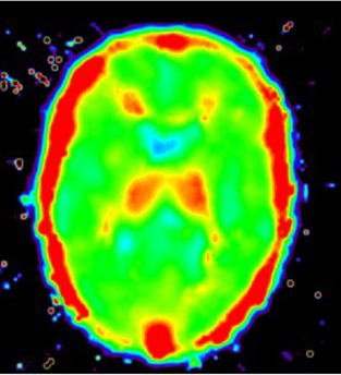
Cost function landscapes revealed that, in general, global minima were located near ‘typical’ curves within this landscape. Occasionally, however, the global minimum was found at large distances from ‘typical’ curves, resulting in unrealistic BP or Vd. From these results a constraint on combinations of basis function was derived (valley of desire) to avoid trapping in global minima outside the physiological realistic range, which could not be achieved by simple tuning the basis function range. Next BP and Vd were calculated using the 2D BFM with and without this constraint. The latter calculations showed that use of this additional constraint reduced BP and Vd bias from 1.27 and 1.12 to 1.20 and 1.05, respectively, at a noise level of 10%. In addition, BP and Vd precision (COV) improved from 78 and 62% to 46 and 15%, respectively. An example of a Vd image showing increased [11C]-(R)-PK11195 binding in the thalamus is given below.
Conclusion
A novel 2D BFM method was developed for generating parametric images of BP and Vd. To avoid large large bias and poor precision at noise levels typical for parametric applications, the method needs to be constrained to specific combinations of basis functions, which can be derived from a typical shape of 2D cost function landscape. Accuracy and precision of this constrained version seems sufficient for calculating parametric Vd images (See Figure 1).