
Introduction
The High Resolution Research Tomograph (HRRT) is a new, dedicated brain PET scanner, designed for a resolution of <3 mm combined with high sensitivity. Although PET is a quantitative imaging technique, many corrections are needed to achieve sufficient accuracy for in vivo applications. Although basic performance characteristics have been investigated 1 , the applicability of this scanner for neuro-imaging, in particular neuroreceptor, applications still needs to be fully assessed. The purpose of this study was to evaluate the accuracy of dead-time, attenuation and scatter corrections, to assess image uniformity and the effect of outside field-of-view (FOV) activity on quantification. To this end, both phantom experiments and a clinical evaluation were performed.
Methods
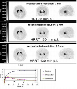
For the experimental study, a 20 cm diameter cylindrical phantom, filled with an 18-F solution with activity concentrations ranging from 1 to 15 kBq/cc, was used. Contrast was created by inserting two cylinders of 5 cm diameter, filled with 1/3 and 2/3 of the background activity concentration. The phantom was measured both with and without a 20 cm cylinder, containing 25 MBq 68-Ge, placed just outside the FOV. Quantitative accuracy was assessed by ROI analysis of the (3D OSEM) reconstructed images. Clinical evaluation included four [18F]-FP-ß-CIT studies. A dynamic 90 minutes scan was acquired on an ECAT EXACT HR+ scanner. After this scan the patient was transferred to the HRRT scanner and an additional scan of 15 minutes was acquired, starting approximately 120 minutes p.i. Time activity curves were generated after alignment of HRRT with HR+ images.
Results
Dead-time and decay corrections were accurate within 1. 0%. Out of FOV activity increased only the randoms rate slightly. Reconstructed activity concentrations in the background and two inserts were within 8% of the expected values, whilst observed contrasts were accurate within 2%. Inter- and intraplane uniformities were 4.3 and 8.3%, respectively. Quantification, uniformity and linearity were not affected by outside FOV activity. Clinical evaluation showed that ROI values of several regions in the HRRT PET images were within 8% of the values extrapolated from HR+ time-activity curves (see figure 1). Furthermore, visual inspection revealed that HRRT images similar to HR+ images, but with superior resolution.
Conclusion
Linearity, dead time and attenuation corrections of the HRRT were sufficiently accurate for clinical applications. Effects of outside FOV activity were minimal, thereby obviating the need for additional shielding. Inter-plane and intra-plane uniformity were, however, suboptimal. Improvements can be expected from new scatter correction methods. Clinical results showed a deviation of less than 8% from HR+ data and images had comparable image quality, but higher resolution. It is concluded that the HRRT is suitable for quantitative neuro-imaging applications, but that it would benefit from refinement of the scatter correction algorithm.