
Introduction
Previous PET studies have suggested that [11C]flumazenil (FMZ) is adequate to map neuronal death in vivo1, 2. Thus, FMZ may be useful for in vivo sequential investigation of neuronal loss following stroke, both acutely in the infarcted tissue and subsequently in the structurally surviving penumbra. To investigate this possibility a small-animal PET scanner (microPET P4) has been used to image central benzodiazepine receptors in the transiently ischaemic rat cortex using FMZ. Six male SHRs have been imaged in a longitudinal study design and FMZ distribution volume (DV) maps have been compared to immunohistology.
Methods
On Day 1 each SHR was subjected to 45 min distal MCAo to maximise cortical ischaemia 3 . Following 60 mins of reperfusion FMZ was injected and image data was acquired for 75 mins. The images were reconstructed using 3D filtered backprojection, with corrections applied for normalisation, randoms, dead time, attenuation, decay and sensitivity. DV maps were calculated from image and input function data using the pmod kinetic modelling package. Further imaging was conducted at 48 hrs and 14dys. After the final scan the experiment was terminated by transcardial perfusion fixation. Brains were processed immunohistochemically.
Results
FMZ binding is significantly lower (p=0.02) in ipsilateral cortex (IC) compared to contralateral cortex (CC) 1 hr after reperfusion. At 48 hrs, subjects with more ischaemic damage (shown immunohistologically) demonstrate higher FMZ binding in IC versus CC. DV values also show a transient global increase in FMZ binding at Day 2. By Day 14, DV maps show lower FMZ binding in IC compared to CC; this effect varied between subjects, depending on the extent of inflammation and neuronal loss observed via immunohistology.
Conclusions
The results indicate bi-phasic changes in FMZ binding in the occluded cortex and globally after temporary brain ischaemia. Final interpretation of the focal FMZ changes will benefit from further histopathological categorisation into infarcted, selective neuronal loss or normal tissue, and from more detailed image analysis. However, other factors, over and above neuronal loss, such as blood brain barrier disruption, endozepine release, and GABAA receptor modulation may be contributing to the observed changes (See Figure 1).
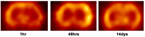
Example subject's DV maps at threee time points post MCA reperfusion. Coronal slices are shown, the ipsilateral hemisphere is displayed on the right.