
Introduction
Rat subarachnoid hemorrhage (SAH) model has been widely used recently but its angiographical assessment is difficult due to small size of the animals to maintain physiological parameters and due to the small size of arterial lumen for angiographic measurement. We developed a microangiography system using monochromatic synchrotron radiation X-rays at SPring-8, a third generation synchrotron radiation facility. In Brain03 meeting we have reported autoregulatory changed of small arteries under hypercapnia and hypotension. In this study, we assessed the distensibility of major trunk arteries after subarachnoid hemorrhage in normotensive and hypertensive rats.
Methods
Twenty adult Wistar Kyoto rats (WKY) and 13 stroke prone spontaneously hypertensive rats (SHR-SP) were prepared SAH by double hemorrhage injection method into cisterna magna or sham operation as control. Microngiography was performed on day 7 by retrograde injection of 0.2 ml contrast media via external carotid artery for imaging of internal carotid artery and 0.4 ml for basilar artery. Angiography was repeated 4 times in each rat before and after loading of hypercapnia at 100–120 mmHg of PaCO2. The diameters of major trunk vessels were assessed. Histological observation of artery lumen and wall were also performed.
Results
Angiographical vasospasm was demonstrated in basilar artery in WKY with 68 % of diameter of control. In ICA and other major trunk in WKY and all the arteries in SHR-SP did not demonstrate vasospasm. Hypercapnia induced loss of distensibility in BA of WKY. In SHR-SP, this distensibility was impaired regardless hemorrhage. Histological study demonstrated basilar artery in WKY thickened at 179 % after SAH and became similar to non-hemorrhagic SHR-SP. ICA in WKY and both BA and ICA in SHR-SP were unchanged in wall thickness before and after SAH.
Conclusions
High quality angiography demonstrated deteriorated distensibility of spastic artery only in basilar artery of normotensive rats, which was well-correlated to histological change of arterial wall. Preexisting pathological deterioration in artery of SHR-SP might lead less affected spastic change after SAH. (See Figure 1)
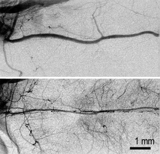
Spastic basilar artery in SHR-SP with SAH before (upper) and after (lower) hypercapnia.