Objective
There has been a series of recent publications using in vitro hippocampal and cortical slices which document changes in cerebral arterioles during neuronal activation. These in vitro vessels studied in slices are non-pressurized and non-perfused. Thus, the appropriateness of extrapolation to the in vivo vascular responses is unclear. The vascular response of isolated pressurized, perfused in vitro cerebral arterioles have been well characterized. In contrast, the responses in non-pressurized, non perfused cerebral arterioles remains to be determined. Consequently, the goal of the following experiments was to study the arteriolar response in non-pressurized, non-perfused cerebral vessels.
Methods
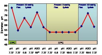
Penetrating intracerebral arterioles from adult male Sprague-Dawley rats were dissected at 4°C and transferred to a temperature controlled bath mounted on inverted microscope. The bath temperature was raised to 37°C and maintained at a constant flow of buffered saline 1 ml per minute. The vessel reactivity was assessed by applying adenosine (ADO) extraluminally at concentration 10E6 M in buffered saline and by changing the pH of the buffered saline from pH 6.8 and 7.6. For comparison, we also determined the responses in pressurized, perfused penetrating arterioles.
Results
We observed minimum or inconsistent reactivity in non-pressurized vessels. There was no significant changes in vessel diameter with warming the bath (from 22°C to 37°C), after application of adenosine at concentration 10E6 M or in response to pH changes. In contrast, in pressurized (60 mm Hg), perfused (2–4 uL) arterioles, we observed vasoconstriction after increase in bath temperature from 22 C to 37 C, vasodilatation in response to adenosine application, and appropriate response to change of the pH of the bath: constriction with raising the pH from 7.37 to 7.6. and vasodilatation from 7.37 to 6.8.
Conclusions
These results signify the importance of intraluminal pressure and flow for preservation of physiological reactivity of penetrating cerebral arterioles. We therefore urge caution in extrapolating physiologic significance of responses observed in non-pressurized, non-perfused arterioles in hippocampal and cortical slices. (See Figure 1).