Introduction
Superparamagnetic iron oxides nanoparticles (SPION) are useful tracking markers of stem cell and used for numerous in vivo applications such as magnetic resonance imaging contrast enhancement. In the present study, we compared three different SPION such as standard superparamagnetic iron oxides (feridex), monocrystalline iron oxide nanoparticles (MION-47), and crosslinked iron oxides with tat peptide (tat-CLIO) in terms of their capacity to label human neural stem cells (hNSCs).
Methods
hNSCs were incubated in cell culture media containing each SPION with which concentrations and incubation times have been modified from previous studies. Cell labeling with iron was assessed by Prussian blue stain and iron contents of the cell were analyzed by atomic absorption furnace spectrophotometer (AAS). Proliferation and vitality of hNSCs after SPION labeling were also evaluated using an investigation of long-term cell proliferation ability to continue and trypan blue dye exclusion test, respectively. The localization of iron oxides in hNSCs was investigated by Prussian blue stain and transmission electron microscopy. Furthermore, the retention of SPION in the cells were evaluated at different incubation time points of 6, 24 and 30 hr.
Results
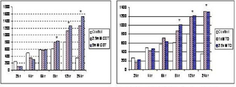
Incorporation of the each SPION appeared not to affect cell proliferation and vitality as compared with these of the nonlabeled cells. Within the cytoplasm iron oxide nanoparticels were presented. Cellular iron was detected in almost all hNSCs that were incubated with feridex or with tat-CLIO, but not with MION-47 as determined by Prussian blue stain. In addition, assessment using AAS revealed that tat-CLIO-labeled cells had more iron contents by two times and ten times when compared with cells labeled with feridex and MION-47, respectively. Moreover, labeling of cells with tat-CLIO revealed the longest retention of SPION in hNSCs up to 30 hr.
Conclusion
Taken together, our study suggests that magnetic labeling of hNSCs using tat-CLIO is a useful tool for tracking and probably magnetic resonance imaging in future stem cell studies. Grant support: supported by a grant (SC13161) from Stem Cell Research Center of the 21st Century Frontier Research Program funded by the Ministry of Science and Technology, Republic of Korea (See Figure 1).