
Introduction
Dysmyelination, caused by defective genes encoding for myelin components is rare in humans (Pelizaeus-Merzbacher disease). Several dysmyelinated animal models have served as a framework to study the potential of cell therapy, in particular as related to the myelinating properties of transplanted neural stem cells, which has also relevance to CNS repair in multiple sclerosis. In vivo magnetic resonance (MR) cell tracking of magnetically labeled cells has been successfully applied to non-invasively visualize the biodistribution and migration of transplanted stem cells and progenitors in two rat models of dysmyelination, the myelin-deficient rat 1 and the shaker rat 2 . In this study, we report on the applicability of serial MRI cell tracking in the dysmyelinated shiverer (shi) mouse brain at high field strength (11.7 T).
Materials and Methods
The LacZ-transfected neural stem cell line C17. 2 was magnetically labeled with Feridex and poly-L-lysine 3 . C17.2 cells (80,000 in 2 μl) (n=9 mice) or an equal volume of saline (n=2 mice) were injected into the right ventricle of neonatal (P1-3) shi mice. In vivo MR imaging was performed at 1, 3, and 5 weeks after cell transplantation using a Bruker 11.7 T Avance spectrometer. Brains were then removed and imaged ex vivo at high-resolution (65 μm isotropic). Cell distribution was verified by staining for myelin, LacZ, dextran, and iron.
Results
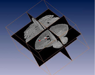
Serial in vivo MR imaging at the different time points revealed that cells appeared to remain within the ventricles for prolonged times. The ex vivo imaging showed migration of hypointense, labeled cells into the brain parenchyma including the corpus callosum, striatum and cortex (Fig. 1). The hypointense signal dots, each representing single cells, closely matched the cell biodistribution as detected by X-gal histochemistry and iron staining. The shaking phenotype of the transplanted shiverer mice, apparent at P12, was not altered throughout the course of 60 days as compared to non-transplanted controls. Consistently, new myelination could not be observed.
Conclusion
These results demonstrate that magnetically labeled neural stem cells migrate long distances from the transplantation site and can be accurately tracked by MR imaging. However, new myelination and rescue of the shiverer phenotype could not be observed. Thus, further optimization of cell therapy will be needed in which MRI cell tracking may become a useful guiding tool.