
Introduction
Elastic image mapping has been applied previously to MRI images to study brain development and various neurological disorders. In this study, elastic mapping is applied to serial MR images of a subject diagnosed with semantic dementia to examine regional brain atrophy associated with the dementia.
Methods
Magnetic resonance imaging (MRI) scans of a 57 year-old male patient diagnosed with semantic dementia were obtained using gradient echo T1-weighted imaging (TR 25 ms, TE 5ms, slice thickness 1.5 mm, FOV 24×18 cm, flip angle 40 degrees, no gaps). A total of four scans were obtained (baseline scan in 02/1993; follow-up scans in 10/1994, 02/1996, and 08/1999). Elastic mapping was then applied to deform later MRI scans (10/1994, 02/1996, and 08/1999) back to the first baseline MRI scan (02/1993). This temporal normalization allows the assessment of tissue expansion/loss in a voxel-by-voxel manner by looking at the determinant of the Jacobian operator (Jacobian map) applied to the resulting deformation field (values larger than 1 indicate tissue expansion; values less than 1 indicate atrophy or tissue loss). A region of interest (ROI) analysis using manual tracing was performed to compute average atrophic rates from the Jacobian maps in regions with observed atrophic changes.
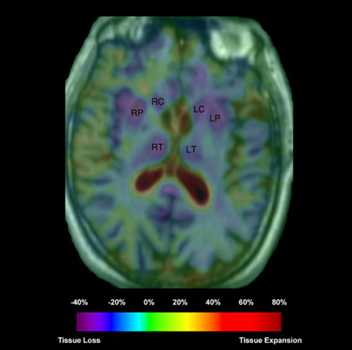
Results
The MRI scans showed existing left temporal lobe atrophy by visual inspection, although no active atrophy was detected in the Jacobian maps during the time period. By contrast, active atrophy was observed in the right temporal lobe. Closer inspection showed a posterior progression of the atrophy starting from the right temporal pole. Both the asymmetrical nature of temporal lobe atrophy and antero-posterior progression are consistent with previous studies. However, in this patient, we also noticed bilateral tissue loss in the caudate head, putamen, and thalamus (Figure 1). An annualized atrophic rate (as a percent change per year of the ROI values) showed more active tissue loss in the right middle and inferior temporal, and occipito-temporal lobe (−2.88%, −3.48%, and −2.88% per year, respectively), as well as bilateral putamen and thalamus (∼−3% per year). Moreover, possible bilateral insula and cingulate gyrus involvement were also noted in the Jacobian maps. Our results showed more extensive brain tissue loss over time in this patient than previously reported by others in the literature.
Conclusion
In addition to atrophy in left temporal lobe, semantic dementia also has progressive right temporal and basal ganglia involvement. Elastic image mapping allows quantitative analysis of serial brain MRI's of semantic dementia patient to show regional tissue loss associated with the progression of the disease.