
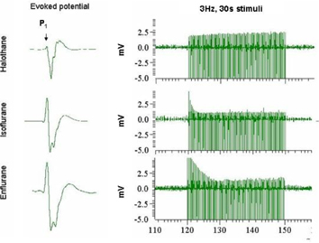
In somatosensory stimulation experiments, non-volatile agents are generally used as the main anesthetic, whereas volatile agents are used for the initial induction phase 1 . This protocol is used because most non-volatile agents themselves do not provide sufficient depth of anesthesia rapidly, which is a specific requirement during the surgical procedure. For the initial surgical preparation volatile anesthetic agents like halothane, isoflurane and enflurane are generally used, because of they are fast acting and have reasonably short half-lifetimes 2 . In this study we examined the effects of these induction agents on artificially ventilated (70% N2O, 30% O2) Sprague-Dawley rats which were anesthetized with á-chloralose (40 mg/kg/hr). We applied strong electrical stimuli at 3 Hz frequency for 30 s period to provoke somatosensory evoked potentials in the forepaw area of the brain approximately 4 hours after stopping exposure to the volatile induction agent. The extracellular recordings were conducted in layer 4 of the somatosensory cortex with Tungsten microelectrodes 3 . The electrical data with high bandwidth (20 kHz) were filtered to obtain field potentials (FP). Since the low pass filtering affects the time course of the electrical signals, we did not examine the latency of the different peaks in the evoked potential. Rather we examined the intensities of the different peaks of the evoked potential (P1, N1, N2, and P2) throughout the stimulation period, for every induction agent (figure 1 left panel). The P1 peak of the evoked potential, which is generally associated with the thalamo-cortical pathway 4 , decreased with time for isoflurane and enflurane induction (figure 1 right panel). On the contrary P1 peak was very much depressed with halothane induction and it was not affected with time during stimulation. Assuming a simple exponential decay in the decrease of the P1 peak we calculated the time constant (where the amplitude decreased to the 1/e level). In the case of isoflurane the time-constant was 7.89±1.04 s (n=8), while the enflurane produced a more elongated decay with a time-constant of 20.86±2.5 s (n=6). The long time-constants with isoflurane and enflurane induction may be a consequence of these agents forming a negative feedback for the thalamo-cortical pathway such that the stimulus-induced evoked potentials adapted over prolonged stimulation 5 . In contrast halothane induction did not influence the adaptive mechanisms. Therefore halothane would seem to be a better choice for an induction agent in neurophysiology measurements (e.g., imaging with optical imaging and fMRI) studies because of the lack of adaptive tendencies in the measured signals during prolonged stimulation figure 1.
Footnotes
Acknowledgements
Supported by NIH (DC-003710, MH-067528) and NSF (DBI-0095173) grants.