
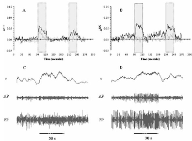
Neuroimaging techniques are predominantly based on changes in CBF induced by alterations in neuronal activity and is generally carried out in animals under anesthesia, usually with α-chloralose because of its minor effects on cardiovascular, respiratory, and reflex functions 1 . General anesthetics reduce neuronal activity in various regions of the mammalian CNS 2 . A considerable number of mechanisms have been suggested to mediate the depressant effects 3 . However it is still a matter of debate as to which molecular targets are truly relevant in producing the “true” anesthetic state 4 . Recent studies reported the relationship between the action of volatile anesthetics occurring on the molecular level and the corresponding effects on neuronal firing 5 . In this study we have used two volatile anesthetics (isoflurane, halothane) and studied the time-dependent induction effects for functional studies in α-chloralose anaesthetized rats. During the animal preparation halothane (0.7%) / isoflurane (0.5%) were used for induction. The same forepaw stimulation protocol (30 s block design; 2 mA; 0.3 ms; 3 Hz) was used for both fMRI and extracellular recordings. All fMRI data (n=10) were obtained on a modified 9.4 T Bruker horizontal-bore spectrometer (Billerica, MA) using a 1 H resonator/surface coil RF probe. The images were aquired with gradient echo EPI sequence (TR/TE=1000/15 ms). Extracellular recordings (n=18) were first filtered to obtain action and field potentials (AP, FP) and then the AP data were binned to obtain spike rates (ν). The changes in local CBF were measured with a laser-Doppler probe (Oxford Optronix, Oxford, UK). The purpose of this study was to examine the effects of isoflurane and halothane used for induction in á-chloralose anaesthetized rats. The fMRI time courses after 3 hrs (gray) and 5 hrs (black) from isoflurane induction (A) showed generally moderate levels of intra-animal reproducibility, which was lacking with halothane (B). These fMRI responses were supported by similar CBF responses measured with laser-Doppler probes (data not shown). The electrophysiology results after 5 hrs (bottom) showed changes in neuronal activity, but the alterations were far less significant with isoflurane (C) than with halothane (D). These results together suggest that while isoflurane induction may result in a faster hemodynamic response, the changes in neuronal activity are slightly depressed. These results suggest caution for interpreting results from anesthetized rats where volatile agents are used for the induction phase (See Figure 1).
Footnotes
Acknowledgements
Supported by NIH (DC-003710, MH-067528) and NSF (DBI-0095173) grants.