
Introduction
Wernicke's encephalopathy (WE), often seen in chronic alcoholics, results from thiamine deficiency and clinically develops opthalmoplegia, ataxic gait, and confusion. It sometimes improves without any defect, but a part of WE subsequently progresses to Wernicke-Kosakoff syndrome (WKS) characterized by continuous amnesia. In chronic WKS, pathological changes are seen mainly in the periventricular regions of the third and forth ventricles and aqueduct where the number of astrocytes is increased but neurons are relatively preserved. Severe neuronal loss occurs in thalamus and inferior olive. An experimental animal model of thiamine deficiency revealed that the reactive activated microglias appear in relatively early stage of WE and it might be a predictor for the prognosis. [11C]PK11195, specific radioligand for peripheral benzodiazepine binding site on activated microglias in the brain in vivo, has been used for imaging neuroinflammation by PET. The purpose of this study is to examine if the activated microglias in WKS can be visualized by [11C]PK11195 and PET as a marker of pathological process to assess status of the disease and prognosis.
Subjects and Methods
We studied seven chronic WKS patients (ages 35–68) and seven healthy volunteers (ages 43–74) who were not habitual alcohol drinkers. All the subjects participated in two PET studies with [18F]FDG and [11C]PK11195. A 6-minute static scan was started at 45 minutes after the intravenous injection of 150MBq [18F]FDG. Regional cerebral glucose metabolism was evaluated semi-quantitatively by normalized regional activity in reference to the global activity. A dynamic PET scan was performed in a 3D mode for 60 minutes after the injection of 500MBq of [11C]PK11195 with serial arterial blood sampling and metabolite analysis. Parametric image of the distribution volume (DV) of [11C]PK11195 was created by Logan-plot method. Volumetric 3D MR images were obtained for anatomical reference. Two PET images were coregistered to individual MRI and regions of interest were placed on thalamus, anterior cingulate, caudate nucleus, and hippocampus. Results The cerebral glucose metabolism was declined in WKS in thalamus, anterior cingulate, frontal and temporal cortex. The DV of [11C]PK11195 was elevated in most of regions in the brain, especially in pons and thalamus (Figure 1).
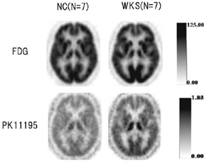
Averaged images of FDG uptake and DV of PK11195 in normals (n = 4) and Wernicke-Korsakoff syndrome patients (n = 3). The elevation of DV in thalamus is shown in this slice.
Conclusions
Our preliminary results suggested that the activated microglias in WKS can be visualized by [11C]PK11195 and PET. This technique may be useful to monitor the pathological process in WKS and to predict the prognosis.