Aim
To establish a bedside method to measure neurovascular coupling (NVC) during functional activation in healthy subjects as well as patients, in whom the NVC may be disturbed, e. g., due to stroke, head trauma or subarachnoid hemorrhage (SAH).
Methods
We investigated the coupling of neuronal activation and cerebral blood flow (CBF) during voluntary hand movement in humans. For this purpose we non-invasively and simultaneously recorded alterations of slow-potential and oxy- and deoxy-hemoglobin (Hb) concentrations over the hand area of the primary motor cortex. Slow potentials were recorded with DC-EEG and oxy- and deoxy-Hb changes with near-infrared spectroscopy (NIRS). 20 female, strongly right handed volunteers underwent a simple motor task consisting of repetitive contraction of one fist for 20 s followed by movement of the other hand for 20 s. Movements were self paced (∼1.5 Hz). Every 20 s a signal was applied (vibrating stimulus at the lower leg) to alternate between hands. We performed 46 stimulation blocks, 23 for each hand so that the whole trial took 15:20 minutes. 15 EEG Ag/AgCl sintered ring electrodes (impedance <2 kW), 8 light sources and 7 light detectors were positioned over the left primary motor cortex with C3 as central recording point. Data processing was performed using BrainAnalyser 1.05 (BrainProducts®) and BESA (MEGIS Software GmbH) for EEG and MatLab 6.5 software (The MathWorks, Inc.) for NIRS data. For spatial correlation, EEG source analysis was related to the point with the strongest decrease in deoxy-Hb. Distance and relation to C3 were determined using a 2D (NIRS) and a 3D (DC-EEG) mapping. Curves of slow potential and the indirect CBF parameters oxy- and deoxy-Hb were then temporally correlated.
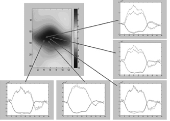
Results
According to the onset of right hand movement we detected a circumscribed negative shift of the DC potential over the left motor cortex accompanied by an increase in the concentration of oxy-Hb and a decrease in deoxy-Hb respectively in the same region. Deoxy-Hb startet to decrease 1.2 s after movement onset and reached its minimum within 6.6 s (t[20%]=2.0 s, t[50%]=3.6 s, t[80%]=4.9 s). Deoxy-Hb increased 2.8 s after cessation of contralateral hand movement. Baseline was reached again within 7.0 s (t[80%]=3.7 s, t[50%]=5.1 s, t[20%]=6.2 s) and an overshoot for 1.6 s was observed subsequently.
Conclusion
We demonstrated that the combination of non-invasive DC-EEG and NIRS imaging is feasible to simultaneously detect changes in DC potential and indirectly in CBF in time and two spatial dimensions. The method is particularly useful to study NVC with a high temporal resolution. We will use the method to study conditions in which neurovascular coupling is likely to be disturbed as in the course of SAH (See Figure 1).