
Background
[11C] flumazenil (FMZ), a ligand that selectively binds to the central benzodiazepine receptor in the neuronal membrane, is useful for evaluating neuronal viability in a PET scan. Using this ligand, we investigated whether there was a correlation between neuronal integrity in various brain structures and vascular parkinsonism (VaP) in patients with leukoaraiosis.
Methods
Twelve patients whose T2-weighted MRI revealed confluent hyperintensities in the subcortical white matter (Schmidt scale score of 3) and several punctate high-intensity areas in the striatum and/or thalamus were studied using PET. Based on a two compartment, two parameter model using metabolite-corrected arterial input and PET-measured cerebral radioactivity, the distribution volume of FMZ (FMZ-Vd) was calculated in various regions of interest (ROIs) by non-linear curve fitting. Additionally, tracer kinetic analysis was applied for voxel-by-voxel quantification of FMZ-Vd and data analysis was performed using statistical parametric mapping (SPM). We also examined cerebral blood flow (CBF) and cerebral metabolic rate of oxygen metabolism (CMRO2) using the 15O gas steady-state method in these patients.
Results
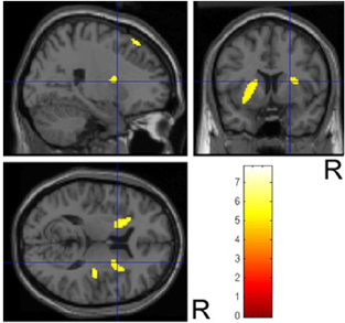
The subjects were divided into two groups based on their neurological signs i.e., patients with parkinsonism (group P; 3 men and 4 women; 73.7+/−4.6 yr) and those without parkinsonism (group NP; 1 man and 4 women; 77.0+/−3.2 yr). The mean+/−SD number of lacunae in the striatum was slightly but not significantly larger in group P (group P, 1.14+/−0.90; group NP, 0.60+/−0.55; p=0.26). ROI-based analysis demonstrated that FMZ-Vd tended to be lower in group P than in group NP. These reductions reached significance in the striatum (17.2%; p<0.01), although no significant difference was found in the other areas. CBF was also reduced in group P, although this reduction only reached significance in the striatum (25.4%; p<0.05). CMRO2 also tended to be lower in group P. The largest reduction in CMRO2 was detected in the striatum (25.3%, p=0.076). SPM analysis showed that FMZ-Vd was significantly reduced in the bilateral striatum and the most rostral part of the right lateral premotor cortex (Puncorr<0.0005; figure 1).
Conclusions
The premotor cortex, striatum, and the frontal white matter interconnecting them are essential components of the basal ganglia-thalamocortical ‘motor’ circuit. Given our findings that neuronal integrity is impaired in the striatum as well as the premotor cortex in VaP patients with WMLs, ischemic damage to the motor circuit may contribute to developing VaP.