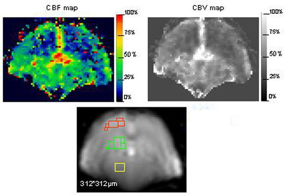
It is generally accepted that knowledge of the regional brain perfusion and assessment of the cerebrovascular reactivity is of great importance in the follow up of neurodegenerative diseases. In particular, since genetically engineered mouse models for neurodegeneration have proven to be of great value for studying the pathogenesis of a disorder at the molecular, cellular and behavioral levels, quantification of cerebral perfusion in these mice becomes extremely relevant. In order to improve in-vivo methods for evaluation of cerebral perfusion in mice, we implemented in-vivo echo planar imaging in control mouse brain and used it for application in bolus tracking (BT) experiments and arterial spin labeling (ASL) acquisitions to assess cerebral perfusion parameters. The BT-technique could quantitatively calculate cerebral blood volume (CBV) and cerebral blood flow (CBF) on a regional basis and ASL was used as a detection method of changing CBF upon a hypoxic challenge. In this way we could identify regional differences in CBV and CBF (Figure 1). The highest CBV values were found in the thalamus (7.59±0.41 ml/100 g). The hippocampus displayed a significant lower CBF at rest conditions as compared to the cortex (67.44±6.44 ml/100 g/min versus 83.17±6.43 ml/100 g/min) and this seemed to be correlated with the observed reduction in the cerebrovascular response to hypoxia (relative CBF increase in cortex 29.41±2.34% versus 15.13±2.29% in the hippocampus) and a more pronounced hypoxia induced BOLD signal change. Our data provide clear evidence that it is possible to discern basal flow parameters in mouse brain and locate differences in spatial patterns of cerebrovascular reactivity in mice. BT-MRI and ASL were proven to provide complementary information on the perfusion status of the brain and this study shows for the first time the advantages of the combined use in mouse brain.