
Objectives
Hyperintensity in diffusion-weighted imaging (DWI) and hypointensity in the apparent water diffusion coefficient (ADC) signals may occur in human stroke 1 , and are seen in animals during MCA occlusion 2 . Yet, these alterations return to baseline levels or partially recover at early reperfusion 3 . Secondary ischemic damage becomes later apparent together with delayed recurrence of the DWI lesions 4 . Here we sought to evaluate whether the degree of early MRI signal intensity changes predicted tissue damage, and to identify the underlying histopathological features.
Methods
Sprague-Dawley rats (n=39) were subjected to transient (60 min) or permanent MCA occlusion and were killed at 8, 12, 15, 18 or 24 h. One or two MRI studies (T2w, DWI) were performed on each rat between 1.5 and 24 h. We evaluated the degree and volume of MRI changes in signal intensity, infarct volume (TTC), histopathology, and immunoreactivity to markers of cellular stress (Hsp72), astrocytes (GFAP), and microglia (OX42).
Results
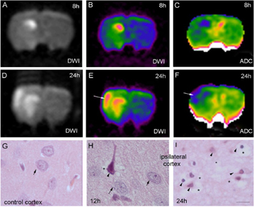
Subtle changes (<15%) in DWI and ADC signal intensity occurred from 90 min to 12 h of reperfusion in the cortex (Figure 1A–C), as compared with greater changes in the striatum, or in both regions after permanent ischemia. The volume of ADC changes at 12 h was predictive of infarct volume at 24 h. These mild changes were correlated with moderate and heterogeneous cell damage (Figure 1H: arrowhead and asterisk; arrows point to normal in G, H). Afterwards, a shift in the degree of cortical MRI signal intensity (>30%) occurred (Figure 1D–F) concomitantly with overt manifestation of the TTC lesion, severe vacuolation of the neuropil (Figure 1I, asterisks), and massive neural cell death (Figure 1I, arrowheads).
Conclusions
Transient ischemia induced a mild histological lesion in the cortex manifesting subtle MRI signal intensity changes up to 12 h of reperfusion. This preceded a high increase in intracellular water content and neural cell death underlying a shift to overt DWI hyperintensity and ADC hypointensity. It is suggested that subtle and persistent MRI changes in the first 12 h hours of reperfusion predict further delayed neuronal death.
Footnotes
Acknowledgements
Supported by CICYT (SAF2002-01963) and FIS. S.R. has a fellowship from MEC.