
Introduction
Benzodiazepine (Bz) receptors may play an important role in anxiety disorders such as posttraumatic stress disorder (PTSD). Indeed, SPECT imaging of Bz binding showed lower distribution volumes in the frontal cortex of patients with PTSD 1 . In addition, rats exposed to acute stress also showed reduced binding to Bz receptors in frontal cortex, hippocampus, and hypothalamus. The objective of the present study was to assess changes in Bz receptor binding in patients with PTSD versus healthy controls using [C-11]-Flumazenil and PET.
Methods
Seven drug naïve veterans with PTSD and seven age matched control veterans with combat experience, but without PTSD, were recruited. Dynamic 3D scans (22 frames) with a total scan duration of 60 minutes were acquired after intravenous injection of ∼370 MBq [C-11]-flumazenil. Arterial blood sampling was performed using both an online detection system and additional manual samples, generating a metabolite corrected arterial plasma input curve. Parametric volume of distribution (Vd) images were generated using Logan plot analysis using the dynamic data from 10 to 60 min p.i. Data were reconstructed using FBP Hanning 0.5, resulting in an image resolution of ∼7 mm FWHM. Furthermore, prior to Logan plot analysis dynamic scans were smoothed using an additional 10 mm FWHM Gaussian filter to reduce noise, thereby avoiding noise induced bias during Logan analysis. Next, these Logan plots were used in a voxel-based comparison between the two groups using Statistical Parametric Mapping (SPM). As images were already smoothed prior to Logan analysis, the usual smoothing within SPM was omitted. SPM was performed both with and without proportional scaling. Proportional scaling may be omitted because Logan plots are quantitatively accurate at the lower noise levels following smoothing.
Results
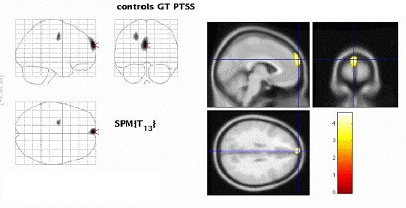
SPM analysis, with and without proportional scaling, revealed no gross differences between patients and controls for most of the brain. However, both analyses, showed a discrete region in the (left) frontal cortex with statistically significantly (p<0.01) reduced [C-11]-Flumazenil binding in PTSD subjects (as indicated by the figure 1).
Conclusions
The observed reduction of [C-11]-Flumazenil binding in the frontal cortex in PTSD subjects is consistent with findings based on SPECT imaging 1 . Other regions did not reveal this reduction, as might have been expected from small animal experiments. Although SPM analysis is useful in identifying regions with changes in ligand binding, full quantification of [C-11]-Flumazenil binding will require further kinetic analysis of the data.