Background and purpose
Since various degrees of hemodynamic and metabolic abnormality and ischemic symptoms are observed in moyamoya disease patients, it is essential to identify the hemodynamic and metabolic status in these patients. In this study, we evaluated the accuracy of quantitative analysis and the ability for detecting misery perfusion by measuring perfusion weighted magnetic resonance imaging (PWI) in moyamoya disease patients.
Methods
Forty-one patients with angiographically defined moyamoya disease were studied with the use of PWI and positron emission tomography (PET) within a month interval of each other. The PWI data were calculated in two analytic methods, one was with deconvolution and the other was without deconvolution. Parametric tomographic axial images to express mean transit time (MTT), cerebral blood flow (CBF) and cerebral blood volume (CBV) was reconstructed. Data obtained on regions of interests (ROIs) among the anterior circulation was relatively expressed by using the data obtained from cerebellum as a control and compared with PET data respectively. The correlation between PWI-MTT and oxygen extraction fraction (OEF) by PET, or PWI-MTT and PET-CBV were also investigated.
Results
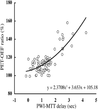
Parametric maps of PWI showed higher resolution than PET map, and indicated focal perfusion failure accurately. From the comparison with PET data, it was revealed that PWI data corresponded well with PET data (CBV, P value was <0.001 in each analytic method, MTT; P value <0.001). On the other hand, there were no significant correlation between PWI and PET data of CBF. From the comparison of PWI-MTT with PET-OEF, in the cerebral hemisphere, which MTT delay compared to cerebellum showed within around 2 seconds, the value of OEF ratio of did not exceed 120%. On the other hand, when MTT delay exceeded 2 seconds, OEF ratio accepted the tendency to begin to go up in proportion to the MTT delay. There was significant correlation between MTT delay and OEF ratio by PET (R=0.755, r2=0.606, P<0.001). This result may suggest that the existence and the grade of misery perfusion would be detectable by measuring PWI.
Conclusion
PWI measurement was sufficient to evaluate CBV and MTT quantitatively in patients with moyamoya disease. Of the various parameters, our results suggested that the degree of MTT delay could possibly be used to detect the existence and degree of misery perfusion (See Figure 1).