
Introduction
Pathologic changes in Alzheimer's disease (AD) typically develop first in the transentorhinal regions of allocortex. The destructive process then spreads to the hippocampus, and eventually encroaches upon the neocortex. A recent study had shown that significant atrophy in the Pcing-Prec was observed in patients with early stage of AD and even in these with presymptomatic stage by using voxel-based morphometry (VBM) technique, which may raise a question as to whether pathological changes in these areas are involved in the early stage of AD. We studied to clarify this question, and have come to advocate a new hypothesis of subtype.
Subjects and Methods
A total of 142 subjects was enrolled in this study. Twenty-six patients with AD and 12 patients with mild cognitive impairment (MCI) were recruited from specialized outpatient units. The diagnosis was based on thorough clinical, neuropsychological, and biological investigations, and all patients fulfilled the NINCDS-ADRDA criteria for probable AD. Since the criteria for amnestic MCI are not yet clearly established, we define MCI as the patients who have memory impairment beyond what is felt to be normal for their age but the MMSE score is over 24. Seventy-four healthy volunteers and 30 young volunteers were selected as control groups. The 3D T1-weighted MR images of the brain were obtained on a 1.5-T Signa Lx with a spoiled gradient-echo technique. For local-level analysis, we employed VBM methodology by SPM99 running on Medx software. We accepted a statistical threshold of p<0.001.
Results
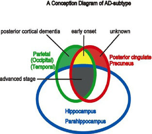
Local decrease of gray matter volume with age was observed bilaterally in the operculum, insula, cingulate (mainly frontal cingulate), caudate nucleus, thalamus (anterior and medial parts), and hippocampus. When comparing the AD group to the age-matched control group, significant volume loss was observed in the parietal, hippocampal, internal orbitofrontal, and Pcing-Prec areas. We also studied the AD patients individually and classified into four groups according to the atrophic patterns; hippocampus, hippocampus plus parietal lobe, hippocampus plus Pcing-Prec area and Pcing-Prec area plus parietal lobe (Fig. 1). The onset age of the last group was significantly younger than the other three groups, but the MMS was not significantly different among these groups. The MCI group was composed of all subtypes except Pcing-Prec plus parietal type.
Discussion
From our results we propose a hypothesis of “4 subtypes” in AD patients. Three of them basically conform to the Braak staging, that is, the initial pathological alterations develop in the transentorhinal and entorhinal regions. During progression of the disease, the affected areas would spread variously to the association cortices in these groups. On the other hand, the Pcing-Prec plus parietal type has distinctive features from the other. Patients in this group show significantly early onset and have relatively little volume loss in the medial temporal area. Atrophic process in the perisylvian and subcortical nuclei seemed to be basically influenced by aging.