Introduction
Cerebral blood volume (CBV) measurements by T2*-weighted dynamic contrast MRI requires an accurate determination of the ratio of gadolinium (Gd) in brain to that in blood. For example, the equation for calculating CBV after a Gd bolus includes the ratio of K(tissue) / K(artery), the proportionality constants relating signal change to Gd concentration in tissue and artery. Although, it is standard to assume that the ratio K(tissue) / K(artery) = 1, this may not be valid since K is dependent on Gd concentration and TE, as well as velocity and orientation. Fortunately, it is not necessary to know either constant but only their ratio. Because tissue and artery proportionality constants relating signal change to concentration for CT are independent of these factors, it can be shown that the ratio of CBV measurements made with identical kinetic paradigms by CT and MRI yields an experimental value for the ratio K(tissue) / K(artery) needed for MRI.
Methods
10 volunteers completed MR and CT perfusion studies within two hours. MR studies (GE 1.5 T Signa) used a bolus (70 μmol/kg Omniscan®, TR1150/TE35, 12 slices, 5 mm) and an infusion (180 μmol/kg over 90 sec, 2500/35, same slices). CT studies (GE CTI ultra-fast 4-slice scanner) also used a bolus (50 ml bolus Omnipaque® at 5 ml/sec, 40 frames, 4 slices, 5 mm) and an infusion (50 ml at 1 ml/sec, same slices, 1sec interscan delay). CBV was calculated for 3 cortical, 4 deep gray and 3 white matter bilateral ROIs on all scans using custom MATLAB software. Area under the curve (AUC) was calculated by both non-parametric (npAUC) and parametric (pAUC, after gamma variate fitting) methods. CT to MRI ratios were determined in each ROI of each subject prior to calculating means or variance.
Results
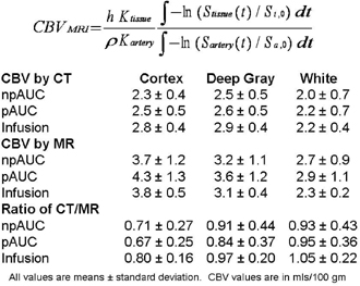
The CT / MRI (and hence K(tissue) / K(artery)) ratios are not different from 1 for any method in deep gray or white matter but are significantly different for cortex. The Infusion method consistently produced the CT / MRI ratios closest to 1, the least variability and the highest gray matter to white matter ratios.
Discussion
The assumption of unity for the proportionality constants is acceptable for deep gray and white matter but not in cortex where “blooming” of signal from leptomeningeal vessels into the parenchyma appears to exaggerate CBV considerably. Measurement of CBV by infusion yields more consistent values than measurement by contrast bolus (See Figure 1).