
Introduction
Peer-reared (PR) rhesus monkeys with early maternal separation later exhibit aggressiveness, impaired impulse control, and alcohol abuse; they also show low CSF 5-HIAA (a serotonin metabolite) and low basal plasma cortisol/ACTH with impaired response to acute stress1, 2. Cortisol facilitates the gene expression of the serotonin transporter (SERT) via the stress axis. However, information on regional 5HT function in vivo in these animals is limited. The purpose was to compare regional brain SERT binding between PR and mother-reared (MR) monkeys with [11C]DASB, a SERT PET tracer.
Methods
Two groups of mean age (3. 4 y) and weight (5.7 kg) matched adolescent male rhesus monkeys (9 PR and 7 MR) were studied. PET data were acquired under isoflurane anesthesia (1.6%) for 2 h with a bolus injection of 4 mCi of [11C]DASB. Additionally, one monkey from each group underwent, on a different day, two 2 h [11C]DASB PET scans separated by 1 h, first with a bolus and infusion (B/I) of high specific activity (SA) (2000 mCi/μmol) and second with B/I of a low SA (μ35 mCi/μmol). From the bolus PET data, parametric images of binding potential (BP=Bmax/KD′, Bmax = transporter density, KD′ = KD/f2, KD = dissociation constant and f2 = free tissue fraction) and relative blood flow (R1) were generated by the two-parameter multilinear reference tissue model using the cerebellum as reference tissue 3 . The parametric images were normalized to a template MRI created from all 16 MRI scans. Group parameter differences were analyzed voxel-wise by a two-sample t test in SPM2. The magnitude of group parameter differences was evaluated by regions of interest (ROIs) analysis. The B/I PET data were used to estimate Bmax and KD′ separately by a Scatchard analysis method.
Results
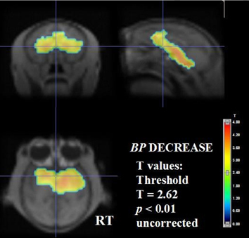
In the PR, BP was decreased (T = 2. 62, p < 0.01) by 15–20% in the raphe, thalamus, striatum, hippocampus/amygdala bilaterally and the remaining right temporal lobe; R1 was also decreased by 15–20% in similar regions more symmetrically except for thalamus where R1 were not different (Fig). The lower striatal BP (0.76) in the PR monkey than that (1.02) in the MR monkey was due to lower Bmax (40 pmol/mL) in the former than that (50 pmol/mL) in the latter with similar KD′ values.
Conclusion
These results agrees with the hypothesis that early maternal deprivation affects the development of the serotonin system and that decreased SERT binding in the critical brain regions may explain some of the behavioural and biochemical abnormalities in PR monkeys.