
Introduction
An abrupt change in brain tissue potassium concentration, [K+]br, has been suggested as an index of progression of ischemic damage 1 . MRI has a potential to study Na+ homeostasis in brain 2 . To monitor K+ by MRI, partial Rb/K replacement has been proposed 3 based on the properties of Rb+ as a congener for K+ in tissues and its much better MR-sensitivity. It is not known, however, to what extent such substitution would influence ion balance in ischemic brain.
Methods
Five normally-fed male Sprague-Dawley rats (240–300 g) were given 30–60 mM RbCl in the drinking water for 12–19 days. Focal cerebral ischemia was induced by MCA transection (MCAT) 4 , and was maintained during 2.5 (n=3) or 5 (n=2) hours. Samples of cortical brain of 1-mm diameter were punched from the ischemic core and contralateral areas 5 using the change in surface reflectivity of ischemic cortex as a guide 6 . Na, K and Rb content in the brain were determined by emission flame photometry at 589, 766 and 791 nm for Na, K and Rb, respectively, and corrected for the mutual interference effects.
Results
The total of [K]br+[Rb]br perfectly fits our previous data on the time course of [K+]br after focal ischemia featuring a sharp drop at 3.5 hours in animals without Rb/K substitution 1 . [K]br and [Rb]br were pooled together, because the degree of Rb/K replacement (i.e., the Rb/(K+Rb) ratio) varied between 0.12 and 0.20 for different animals. As expected, [Na+]br concentration increased with time after ischemia onset. The Rb/(K+Rb) ratio increased over time in ischemic areas (normalized to the contralateral brain): 1.057±0.007 at 2.5 hours and 1.15±0.01 after 5 hours post MCAT (p<0.05). This suggests that the Rb+ efflux from the injured tissue is slower than the K+ efflux.
Conclusion
Rb+ loading does not significantly change the pattern of [K+]br decrease in the ischemic brain over time. Therefore, the animals with partial Rb/K substitution are suitable for 87Rb MRI studies of K+ homeostasis in ischemia (See Figure 1).
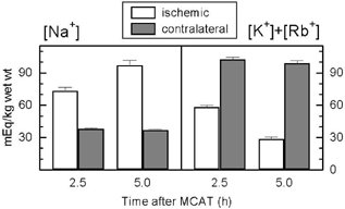
Concentrations of monovalent cations (average±SEM over all animals in the corresponding time groups) in the rat brain cortex after MCAT.
Footnotes
Acknowledgements
Support: NIH grant R01 NS 30839