We designed a mouse model of chronic cerebral hypoperfusion, which invariably exhibits white matter lesions. In the present study, we present histological and behavioral changes after chronic cerebral hypoperfusion in the mouse, and compare the advantages and disadvantages between the rat and mouse models. Application of this model to knockout or transgenic strategies is also discussed.
Methods
Chronic cerebral hypoperfusion was induced by applying microcoils with diameters of 0. 18 mm to the bilateral common carotid arteries (CCAs) in the mouse. Rat model was prepared by clipping the bilateral CCAs. The mouse model was subjected to a set of behavioral assessment tests; rotarod test, open field, light/dark transition, prepulse inhibition, Porsolt forced swim and 8-arm radial maze test. Astroglia and microglia were examined with immunohistochemistry for glial fibrillary acidic protein (GFAP) and MHC class I antigen, respectively. The severity of white matter lesions was graded in myelin staining. Since matrix metaloproteinase 2 (MMP2) is known to be involved in the pathogenesis of white matter lesions, chronic cerebral hypoperfusion was applied to the MMP2 (−/−) mice and wild type mice.
Results
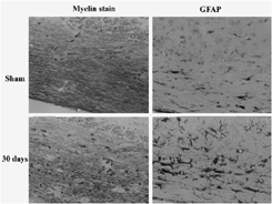
Until 14 days after the operation, CBF ranged between 32–59% in the rats and 69–81% in the mice. Astroglia and microglia were markedly proliferated in the white matter of the both species after chronic cerebral hypoperfusion. White matter rarefaction developed after 14 days in the rats and after 30 days in the mice. Degeneration of the visual pathway was observed exclusively in the rats. Among the set of behavioral assessment scales, the mouse model showed a significant abnormality only in 8-arm radial maze test. Chronic cerebral hypoperfusion induced marked rarefaction of the white matter and activation of glial cells in wild type mice, but not in MMP2 (−/−) mice.
Discussion
We successfully developed a mouse model of chronic cerebral hypoperfusion, which shows cognitive abnormalities without a significant damage to the visual system. Although behavioral abnormalities in the rat model have been reported previously, we did not examine the behavioral tests, since degeneration of the visual system seem to compromise the results. The rat model is more suitable for drug evaluation, because of more prompt emergence of white matter lesions and availability of osmotic minipump. The mouse model is easily applicable to genetically modified animals and also has advantages over the rat model in cognitive evaluation ().