
Introduction
Pathological changes in brain tissue during and after stroke may involve injury to the blood-brain barrier (BBB). In this study, changes in BBB permeability in ischemic tissue were assessed in a rat model of transient focal ischemia that produces BBB injury acutely and hemorrhagic transformation at 24 hours. Changes in the blood-to-brain transfer constant (Ki) were measured by both magnetic resonance imaging (MRI) and quantitative autoradiographic (QAR) methods using identical non-labeled and radiolabeled gadolinium-diethylenetriaminepentaacetic acid (Gd-DTPA) preparations, respectively.
Methods
Transient ischemia was induced in male Wistar rats (n=5) by intraluminal suture occlusion of the middle cerebral artery and withdrawal of the occluding filament after 3 hrs. MRI studies were performed at 7 Tesla. Quantitative MRI assessment of ischemia induced BBB permeability changes was performed approximately 2 hrs after reperfusion using Look-Locker based T1–weighted imaging to generate estimates of Ki via the Patlak plot methodology 1 . MRI localization of areas with BBB opening was performed by tracking contrast enhancement changes produced by Gd-DTPA administration using a Look-Locker T1 sequence. The Gd-DTPA injection was delivered using a stepped down continuous i.v. infusion protocol that maintained a relatively constant plasma Gd-DTPA concentration for approximately 20 minutes. After MRI, the rats were infused with 14C-labeled Gd-DTPA using the same infusion procedure, and then sacrificed for QAR. Both versions of the Gd-DTPA preparation were homemade and identically prepared. Tissue sections were prepared for QAR for confirmation of BBB leakage areas and to provide quantitative blood-to-brain transfer constant (Ki-QAR) estimates. The ischemic area regions of interest (ROIs) with BBB opening were segmented from normal tissue using ISODATA segmentation 2 in conjunction with the serially acquired Look-Locker T1 maps. The MRI defined ROIs were superimposed onto the 14C-labeled Gd-DTPA autoradiograms for measurements of BBB permeability.
Results
Acute BBB disruption was detected in the preoptic area and/or striatum in all rats by Gd-DTPA enhanced MRI and corresponded closely with areas identified by 14C-Gd-DTPA QAR. Estimates of Ki by both MRI and QAR methods were significantly elevated after reperfusion as compared to brain regions with intact BBB function. A scatterplot of the MRI versus QAR estimates of Ki, for the non-labeled and radiolabeled Gd-DTPA are shown in the Figure 1. Post-reperfusion BBB changes between MRI and QAR were highly correlated in areas with BBB disruption (p=0.002).
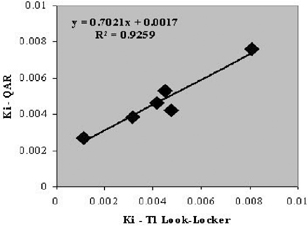
Scatterplot plot of Ki estimates as measured by MRI using Tl estimates plotted as a function of Ki estimates as measured by QAR methods.
Conclusion
The correlation of blood-to-brain transfer constants for non-labeled and radiolabeled Gd-DTPA preparations using a continuous infusion schedule for both MRI and QAR methods provides a one-to-one validation of the Patlak methodology for estimating BBB permeability in reperfused ischemic infarct. With further substantiation, this approach may have potential application in the clinical setting for assessing acute BBB injury that may precede later hemorrhagic transformation.
Footnotes
Acknowledgements
Grant support: Supported in part by NIH RO1NS38540 and AHA 0270176N