
Introduction
Acute loss of function and subsequent spontaneous recovery after stroke have been associated with alterations in neuronal connections. Manganese-enhanced MRI allows in vivo mapping of disruption of neuronal tracts after stroke 1 , however the temporal pattern of manganese enhancement has not yet been fully characterized. The aim of this study was to track changes in connectivity within the sensorimotor network in rat brain after stroke by repeated manganese-enhanced MRI measurements.
Methods
Transient focal cerebral ischemia was induced in six male Wistar rats by 90 minutes intraluminal occlusion of the right middle cerebral artery. After 2 weeks, 0.2 ml 1 M MnCl2 solution was injected into the spared ipsilesional sensorimotor cortex. MnCl2 was also injected in the sensorimotor cortex of six control rats. MRI measurements were done at 2 days before and at 6 h, 24 h, 2 days, 4 days, 6 days and 8 days after MnCl2 injection. T2-weighted images were acquired for lesion assessment and anatomical details. T1-weighted MRI was performed to quantify T1 shortening by manganese in four ipsi- and contralateral regions-of-interest (ROIs) within the sensorimotor network (i.e., sensorimotor cortex (SMCX), caudate putamen (CPu), thalamus (Th) and substantia nigra (SN)). Data were statistically analyzed using a two-way repeated measures ANOVA with a post-hoc Bonferroni t-test.
Results
In all rats, the cortico-striatal-nigral pathway was identified by T1 shortening after MnCl2 injection (Fig 1). In stroke rat brains, however, T1 shortening was significantly reduced in the ipsilateral thalamus and substantia nigra, as compared to control rat brains (P < 0.05). No significant differences were found for other ROIs. Maximal T1 shortening was observed at day 2 or 4 after manganese injection. T1 shortening was significantly reduced after 6 and 8 days as compared to the time-point of maximal T1 shortening in all ROIs except for the ipsilesional thalamus and substantia nigra in stroke rats.
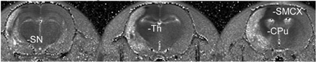
T1 maps of 3 adjacent coronal brain slices of an ischemic rat brain 2 days after injection of manganese chloride. The ischemic lesion is characterized by a prolonged T1.
Discussion
Different spatial and temporal pattern of manganese enhancement in ipsilesional sensorimotor network after stroke suggests disturbed neuronal connections and transport dynamics. MRI mapping of the spatio-temporal distribution of the neuronal tracer manganese can provide unique in vivo information on neuronal connectivity that may aid in elucidating mechanisms of functional loss and recovery after stroke.