
Background
Neurocan is one of the major chondroitin sulfate proteoglycans in the nervous tissues, whose expression and proteolytic cleavage are developmentally regulated. Full-length neurocan (275 kD) is cleaved to yield C-terminal (150 kD) and N-terminal (130 kD) fragments in the adult brain. Both full-length and N-terminal neurocan inhibit axonal extension or regeneration, indicating their important roles in the brain after ischemia. In the present study, therefore, we investigated the expression of neurocan in the brain after ischemia.
Methods
We used male Wistar rat of 12-weeks old. Under the anesthesia with nitrous oxide and halothane, the origin of the right middle cerebral artery (MCA) was occluded using a nylon thread. Ninety minutes later, the cerebral blood flow (CBF) was restored by removal of the thread. The animal was decapitated at 1, 2, 4, 10 and 20 days after the reperfusion and the brain was used for the immunohistochemical and Western blotting analysis as described below. The distribution of neurocan overexpression was investigated by immunohistochemical analysis. We carried out double staining of neurocan and glial fibrillary acidic protein (GFAP) in order to investigate the relationship between neurocan expression and reactive astrocytes. The temporal profile of neurocan expression was investigated by Western blot analysis (n=3 for each time point).
Results
Immunohistochemical analysis showed that a strong signal for neurocan appeared 4 days after reperfusion in the peri-ischemic region of cerebral cortex and caudate (Fig. 1). Double fluorescence study showed that many GFAP-positive cells existed in the area of strong neurocan expression, indicating that neurocan should be produced by reactive astrocytes. Western blot analysis showed that the full-length neurocan appeared in the peri-ischemic region from 1 to 20 days after reperfusion with a peak at 4 days (Fig. 2).
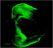
Immunohisto chemistry for neurocan
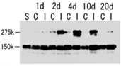
Western blot analysis for full-length (275 kD) and C−terminal (150 kD)neurocan S, sham operated C, contralateral cortex I, ipsilateral cortex
Conclusion
Full-length neurocan was increased at the peri-ischemic region of ischemic brain. Accumulation of the full-length neurocan produced by reactive astrocytes may be one of the processes for tissue repair and reconstruction of neural networks after focal brain ischemia as well.