
Introduction
Increased levels of TGF-ß1 have been implicated in the cerebrovascular dysfunctions that accompany Alzheimer's disease. Transgenic mice that overexpress TGF-ß1 exhibit perivascular accumulation of amyloid and basement membrane proteins, thinner endothelial cells, decreased local cerebral blood flow (Buckwalter et al., 2002, Ann NY Acad Sci 997, 87) and, as we recently found in middle cerebral arteries (MCAs) from such aged mice, a reduced contractile response to ET-1 (Tong et al., submitted). As ET-1 is an important regulator of cerebrovascular tone and homeostasis, we tested if chronic overproduction of TGF-ß1 could decrease contraction to ET-1 via inactivation of its signal transduction pathway.
Methods
ET-1-induced contractions were measured in isolated and pressurized MCAs from aged (p>16 months) TGF-ß1 transgenic and wild-type littermate controls (n=3/group), or young C57Bl/6 (n=10) mice in the presence or absence of 25μM SB-203580 (p38 MAPK inhibitor) or 10μM U0126 (extracellular signal-regulated kinase (ERK1/2) kinase or MEK inhibitor). Primary cultures of rat brain microvascular smooth muscle (SMC) cells were generated from 112 μm mesh-harvested cortical microvessels. Cells were exposed (3 days) to TGF-ß1 (3 ng/ml) in the absence of serum, and then stimulated with ET-1 (10–7 M, 10 min) with or without pre-incubation (1 hr) with SB-203580 (25 μM) or U0126 (10 μM). Alternatively, cells were exposed to TGF-ß1 (2 days) and, on the 3rd day, to both TGF-ß1 and Ro-31-8220 (2–6 μM, an inhibitor of MKP-1 expression), before ET-1 stimulation. Cell lysates were separated by SDS-PAGE, transferred to nitrocellulose filters, probed with antibodies against ETA and ETB receptors, MKP-1, cyclo-oxygenase-2 (COX-2, marker of cell activation) or phosphorylated-MKK3/6, -ERK1/2, or -p38 MAPK.
Results
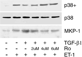
MCAs from aged TGF-ß1 transgenic mice had reduced contractions to ET-1 as compared to wild-type littermates (39%, p<0.01). Similarly, MCAs from young mice treated with SB-203580 showed severely impaired contractions to ET-1 (68%, p<0.001) while U0126 exerted a much smaller effect (32%, ns), indicating that p38 MAPK is the main transduction pathway in this response. In SMC cells, chronic exposure to TGF-ß1 increased COX-2 expression, had no effect on levels of ETA and ETB receptors, MKK3/6, but significantly decreased (18.5%, p<0.05) phosphorylated p38 MAPK levels while increasing those of MKP-1 protein, indicating selective inhibition of the down-stream ET-1 transduction pathway at the p38 phosphorylation step. Further, SMC cells treated with TGF-ß1 and Ro-31-8220 showed normalized levels of phosphorylated p38 MAPK and decreased MKP-1 protein levels.
Conclusion
These results demonstrate that chronic high levels of TGF-ß1 induce MKP-1 expression in cerebrovascular SMC cells, which leads to the inactivation of p38 MAPK and altered capacity of the brain vessels to constrict in response to ET-1. TGF-ß1 may thus exert detrimental effects on cerebrovascular tone and homeostasis by promoting MKP-1, a negative regulator of p38 MAPK activity (See Figure 1).
Footnotes
Acknowledgements
Supported by CIHR (MOP-64194) and Alzheimer Society of Canada.