




Introduction
Microcirculatory function of the brain is essential for providing adequate oxygen to the tissue cells. In neurovascular diseases disturbances of the cerebral microcirculation possibly plays a role in the development of cerebral ischemia. Until recently, the in-vivo observation and quantitative functional assessment of the human cerebral microcirculation were limited by the absence of appropriate investigational techniques. This limitation have been overcome with the introduction of orthogonal polarization spectral (OPS) imaging. We used OPS imaging on the brain cortex during aneurysm surgery and resection of an arterio-venous malformation (AVM) to directly observe the small cortical blood vessels and quantify the contractile properties and changes in microvascular flow (MFI) and functional capillary density (FCD).
Method
In 22 patients undergoing aneurysm surgery the diameter changes of small cortical vessels (15 – 180 μm) were observed using OPS imaging. Fifteen patients were operated early (within 48 hours after bleeding) and 7 underwent late surgery. Immediately after dura opening, the response to hyperventilation (n=16) and papaverin (n=6) of arterioles and venules was observed with OPS imaging under sevoflurane anesthesia. Furthermore, images of the cerebral microcirculation were obtained in two patients undergoing microsurgical removal of an AVM. Eight patients undergoing craniotomy for a disease not affecting the cortical microcirculation served as controls.
Results
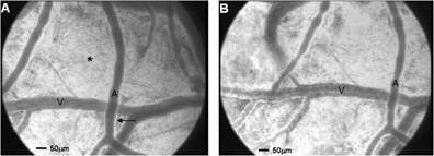
In the presence of subarachnoid blood a severe disturbed reaction of the microvessels was observed. More specifically, hyperventilation resulted in a 39 ± 15% (p<0.05) decrease in arteriolar diameter with a “bead string” constriction pattern occurring in 60% of patients (figure 1a and 1b). The topical application of papaverin resulted in an increase in arteriolar diameter of > 50%. These reactions were not seen in the absence of subarachnoid blood (late surgical patients and controls). In the cerebral cortex surrounding an AVM, MFI and the FCD were decreased to 2 (moderate flow) respectively 1.4 ± 1.3 cm/ mm2. After AVM excision a substantial increase in MFI to 4 (high flow) was seen accompanied by an increase in FCD to 2.1 ± 0.8 cm/ mm2.
Conclusions
OPS imaging allows direct in vivo observation and quantitative functional assessment of the cerebral microcirculation. Thus, microvascular reactivity and hemodynamic changes in the human brain at the microcirculatory level can be readily assessed and correlated with metabolic and clinical parameters. This new tool may contribute to a better understanding of the pathophysiology of cerebral ischemia.