
Object
The double hemorrhage model in the rat is widely accepted to represent a reasonable simulation of delayed cerebral vasospasm (CVS) after subarachnoid hemorrhage (SAH). A neurological and angiographic characterization of this CVS, however, was not available so far and is provided in the present investigation. Additionally, perfusion weighted imaging (PWI) at 3 tesla magnet resonance (MR) tomography was implemented for determination of cerebral blood flow (CBF) and volume (CBV).
Methods
In a prospective experimental setting CVS was induced by application of 0. 2 ml of autologous blood in the Cisterna magna done twice. Surviving rats were examined on the days 2, 3, 5, 7 and 9. Animals were neurologically graded between 0 and 3, followed by MRI, and selective digital subtraction angiography (DSA). The relative regional (rr) CBF and rrCBV was set in relation to the perfusion (MBF) and the blood volume of the masseter muscle (MBV).
Results
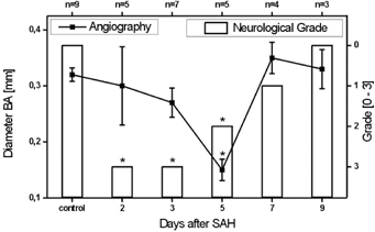
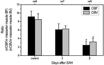
Neurological condition was significantly reduced on day 2, 3, and 5 (medians) (Figure 1). Basilar artery (BA) diameter, however, was not significantly reduced until day 5 reaching 0.15±0.02 mm (SAH) versus 0.32±0.01 mm (sham) (mean ± SEM). In correlation the rrCBF/rrMBF ratio (2.5±0.8 (SAH); 9.2±1.3 (sham)) and the rrCBV/rrMBV (3.2±0.5 (SAH); 8.5±1.3 (sham)) was also significantly decreased on day 5 (Figure 2). Correlation between BA diameter and rrCBF/rrMBF was 0.70 and 0.77 for rrCBV/rrMBV.
Conclusion
A valid and reproducible CVS in the rat double hemorrhage model was proven by neurological score, DSA, and PWI on day 5. Additionally, our data demonstrate the practicability and validity of MR PWI for the monitoring of CVS in a rat SAH model.