
Object
For preoperative studies in cases of carotid endarterectomy (CEA), conventional arterial angiography has been used as a golden standard to evaluate extent of carotid stenosis. Three-dimensional computed tomography angiography (3D-CTA) has been developed and utilized in visualization of various cerebral lesions. Here we present a preliminary report of our experience with CEA using perioperative 3D-CTA in place of selective angiography for evaluation of carotid stenosis.
Methods
A total of 62 carotid arteries were examined before and after CEA, 26 with an early 3D-CT system and 36 with a multidetector helical CT allowing sophisticated reconstruction by personal workstation. In addition to patients who had undergone conventional angiography at other institutes, ten subjects underwent CEA on the basis of 3D-CTA findings alone.
Results
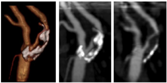
Three D-CTA provided detailed information with an excellent view of carotid stenoses. Volume rendering images comprehensively visualized lesions and surrounding structures as well as calcifications, which were also well depicted by maximum intensity projection images (Fig. 1). With postoperative evaluation, amelioration of stenosis of carotid arteries and disappearance of calcifications on walls were obviously displayed in 3D images. Evaluation of the cerebral circulation is one problem that still requires solution, although cerebral vessels were delineated by 3D-CTA. One patient experienced transient hemiparesis but no significant permanent deficit.
Conclusions
Our preliminary experience of CEA on the basis of 3D-CTA without conventional angiography indicates that this approach with a high-performance workstation provides detailed images with satisfactory preoperative information for CEA. We conclude that 3D-CTA is a safe and accurate modality which is a practical alternative to conventional perioperative angiography.