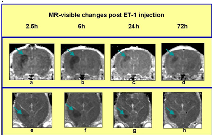
Ischaemic brain events necessitate prompt diagnosis and clinical intervention if the ischaemia is to be reversed. At present, MR is unable to distinguish between reversibly and irreversibly damaged tissue. Although apparent diffusion coefficient (ADC) MRI is established as a sensitive indicator of acute ischaemia, an unresolved challenge is to non-invasively distinguish between reversibly and irreversibly damaged tissue early in the ischaemic event. Endothelin-1 (ET-1) produces a potent and prolonged vasoconstrictive effect on the brain vasculature and has been used as a model of low flow ischaemia in the rat brain. We hypothesise that a focal striatal injection of ET-1 creates a graduated lesion consisting of both reversibly and irreversibly damaged tissue. A longitudinal study of MR-visible changes following intrastriatal ET-1 injection in the juvenile rat brain was conducted and correlated with the metabolic status of brain tissue using phosphorus (31P) MRS. Male Wistar rats (50–55 g) were anaesthetised by inhalation using 2% isoflurane in 70% N2O:30% O2. Using a minimally-invasive stereotaxic procedure, 160pmoles of ET-1 was injected in a volume of 1.0 μl of sterile saline into the striatum (n=8). Animals were positioned in a purpose-built head restraint system and a 2 cm-diameter single-turn circular surface phosphorus coil was placed over the animal's head. Anaesthesia was maintained in the magnet using 1% isoflurane in 70% N2O:30% O2. T1 and T2-weighted images, DWI and 31P spectra were obtained using a 7 T horizontal bore magnet (Magnex Scientific) interfaced to a Varian/SISCO spectrometer (Varian, Palo Alto, CA, USA). We observed a significant decrease in the ADC around the site of injection at 2.5 h post-injection [Figure 1a & e]. MRI was repeated at 6, 24 and 72 hrs over which time ADC changes resolved in the periphery of the lesion, but remained low at the core. These findings suggest that the tissue at the core of the lesion may be irreversibly damaged whereas normalisation of ADC in the periphery may be indicative of reversibly damaged tissue. 31P spectra from the whole of the injected hemisphere did not detect any reductions in the PCr to ATP ratio. Significant changes in these metabolites may only occur in the speculated irreversibly damaged core of the lesion, which constitutes less than 25% of the 31P MRS voxel. The inclusion of a significant volume of healthy tissue in the 31P MRS voxel may explain the absence of a change in metabolite levels. Ongoing studies are investigating the perfusion changes in the core and peripheral regions of ET-1-induced focal lesions and their correlation with ADC changes.