
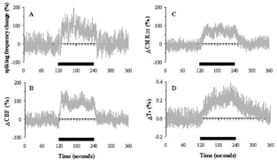
Maintaining near constant brain temperature (Tt) over a wide range of metabolic activity is critical for normal brain function 1 . However brain temperature is an often ignored parameter in neuroscience and clinical studies. Time-dependent variations in Tt are likely to be caused by fluctuations of CBF and CMRO2, both of which are seemingly coupled to alterations in neuronal activity. To this end, we combined magnetic resonance, optical imaging, temperature sensing, and electrophysiological methods in α-chloralose anesthetized rats to obtain multi-modal measurements during forepaw stimulation. Localized changes in neuronal activity were co-localized with regional increases in Tt (by ∼0.2%), CBF (by ∼95%), and CMRO2 (by ∼73%). The time-to-peak for Tt (42±11 s) was significantly longer than those for CBF and CMRO2 (5±2 and 18±4 s, respectively). Net heat (Qnet) in the region of interest (ROI) was modeled as being dependent on the sum of heats attributed to changes in CMRO2 (Qm) and CBF (Qf) as well as conductive heat loss from the ROI to neighboring regions (Qc) and to the environment (Qe). Although tissue cooling due to Qf and Qc can occur and are enhanced during activation, the net increase in Tt corresponded to a large rise in Qm, whereas effects of Qe can be ignored. The study shows that Tt increases slowly (by ∼0.1 C) during physiologic stimulation in α-chloralose anesthetized rats. Since the potential cooling effect of CBF depends on the temperature of blood entering the brain, Tt is mainly affected by CMRO2 during functional challenges. While the importance of the “uncoupling” between changes in CMRO2 and CBF and the temperature of arterial blood for the cooling efficiency of the brain are obvious, our current experimental and theoretical results reveal the significance of the resting CBF and more importantly resting CMRO2 for temperature regulation in the brain. Although the changes in brain temperature recorded for the entire study were spread over a wide range, the stimulation-induced change of ∼0.1 C in the ROI was significant for the majority of the experiments. The small changes in Tt were reproducibly measured from layer 4 of the rat's somatosensory cortex where the highest neural activity changes (induced by forepaw stimulation) were localized. The current temperature measurements from the brain are in accord with previous functional studies in animals2, 3. This is the first study which provides a neuroenergetic basis of regional temperature changes in the brain. The results have implications for functional studies and temperature regulation. (See Figure 1).
Footnotes
Acknowledgements
Supported by NIH (DC-003710, MH-067528) and NSF (DBI-0095173) grants.