
Background
Glucose is the major energy source the adult brain utilizes under physiological conditions. Recent findings, however, have suggested that neurons obtain most of their energy from the oxidation of extracellular lactate derived from astroglial metabolism of glucose transported into the brain from the blood. In the present studies we have used a fluorescent analogue of 2-deoxyglucose, which is often used to trace glucose utilization in neural tissues, to examine glucose metabolism in neurons in vivo.
Methods
Utility of 2-[N-(7-nitrobenz-2-oxa-1,3-diazol-4-yl)amino]-2-deoxy-D-glucose (2-NBDG) to evaluate glucose metabolism was assessed through in vitro study. We incubated cultured neurons and astroglia with 2-NBDG for predetermined time and washed unmetabolized dye out. Phosphorylated 2-NBDG remaining in the cells was measured with a fluorescent microscope, a CCD camera, and an image analysis system. To determine glucose utilization in neurons in vivo, Sprague-Dawley rats were intravenously injected with a pulse bolus of 2-NBDG and decapitated 45 minutes later. After recording the distribution of phosphorylated 2-NBDG, the brain section was further stained immunohistologically.
Results
In neurons as well as in astroglia, fluorescence intensity increased proportionately to incubation time (fig 1). These findings reconfirmed previous in vitro reports that both types of brain cells utilize glucose as their energy source 1 . Phosphorylation of 2-NBDG was linearly dose-dependent up to 90 micro-M (fig 2), suggesting that hexokinase which phosphorylates glucose was not saturated with the tracer at these concentrations. The metabolized dye remained in the cells at least 60 min after wash. The finding is in good agreement with previous reports that both neurons and astroglia contain negligible, if any, amount of glucose-6-phosphatase 2 , an important fact when measuring glucose utilization in the brain using 2-deoxyglucose. These results indicated that, similar to 2-deoxyglucose, 2-NBDG is useful to determine glucose uptake in the brain cells. Examination of hippocampus sections of rats revealed that phosphorylated 2-NBDG accumulated in anti-NeuN positive neurons in the pyramidal cell layer and that GFAP-positive astroglial contribution to overall 2-NBDG accumulation was minor. Sagittal sections of the cerebellum demonstrated that 2-NBDG accumulated in molecular layer, Purkinje cell layer and granular cell layer. Immunostaining for calbindin showed that Purkinje cells in the section all contained metabolized 2-NBDG. Sections at the parietal cortex also suggested the 2-NBDG phosphorylation in neurons as well as astroglia but we could not identify the cells in these sections.
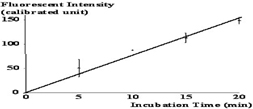
Phosphorylation of 2-NBDG in neurons.
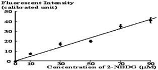
Phosphorylation of 2-NBDG in astroglia
Conclusion
This is the first study demonstrating in vivo glucose uptake and metabolism in neurons in rat brain under physiological conditions. Even though astroglial contribution to overall glucose consumption seemed minor, further studies are needed to confirm the extent of glucose utilization compared to lactate in neurons in vivo.