
Abstract
Cerebral ischemia in the territory of the middle cerebral artery (MCA) can induce delayed neuronal cell death in the ipsilateral substantia nigra (SN) remote from the primary ischemic lesion. This exofocal postischemic neuronal degeneration (EPND) may worsen stroke outcomes. However, the mechanisms leading to EPND are poorly understood. Here, we studied the time course of EPND via sequential magnetic resonance imaging (MRI) and immunohistochemistry for up to 28 days after 30 minutes occlusion of the MCA (MCAo) and reperfusion in the mouse. Furthermore, the effects of delayed treatment with FK506 and MK-801 on the development of EPND were investigated. Secondary neuronal degeneration in the SN occurred within the first week after MCAo and was characterized by a marked neuronal cell loss on histology. Sequential neuroimaging examinations revealed transient MRI changes, which were detectable as early as day 4 after MCAo and thus heralding histologic evidence of EPND. Treatment with MK-801, an established anti-excitotoxic agent, conferred protection against EPND even when initiated days after the initial ischemic event, which was not evident with FK506. Our findings define a secondary time window for delayed neuroprotection after stroke, which may provide a promising target for the development of novel therapies.
INTRODUCTION
Stroke is among the principal causes of long-term disability and death worldwide. 1 To date, treatment options are still very limited, in particular due to the narrow time window for thrombolysis.2–4 Cerebral ischemia in the territory of the middle cerebral artery (MCA) may induce delayed neuronal cell death in non-ischemic brain regions remote from the primary lesion with histopathologic changes occurring as late as days to weeks after the initial ischemic event. 5 This phenomenon, also termed ‘exofocal postischemic neuronal death (EPND)’ has been associated with impaired neurologic function6,7 and the development of vascular Parkinson's disease.8,9 We hypothesize that, due to its delayed occurrence, EPND could also be a promising target for new neuroprotective strategies beyond the narrow time window for acute stroke treatment. However, so far, the mechanisms and characteristics of EPND have remained incompletely understood. 7 In particular, an examination of the time course of secondary changes in a mouse model of stroke is indispensable for studying new treatment approaches. Therefore, we here investigated the pathogenesis of EPND via sequential magnetic resonance imaging (MRI) and immunohistochemistry in a well-established murine model of mild transient brain ischemia. To further elucidate potential pathophysiologic mechanisms involved, we tested the impact of MK-801, an established anti-excitotoxic agent, and of FK506, a potent anti-inflammatory compound. 10
MATERIALS AND METHODS
Animals and Treatments
All experimental procedures were approved by the respective official committee (G0383/09, LaGeSo, Berlin, Germany) and performed in accordance with the Animal Welfare Act, the European Communities Council Directive of 24 November 1986 (86/609/EEC) and the ARRIVE (Animals in Research: Reporting
The study included a total of 68 animals that were subjected to MCAo. Six animals were excluded due to insufficient drop of relative cerebral blood flow and lack of infarct on MRI (please see below). Separate groups of animals were used for histologic characterization, the MRI time course, and the evaluation of drug treatments. Starting on day 5 after MCAo, subsets of animals (
Model of Cerebral Ischemia
Mice were anesthetized by 1.0% (vol/vol) isoflurane in 69% nitrous oxide (N2O) and 30% oxygen (O2) with a facemask. Focal cerebral ischemia was induced by 30 minutes filamentous MCA occlusion (MCAo) and reperfusion as described previously.11,12 Rectal temperature was controlled and kept constant at 36.5±0.5°. Regional cerebral blood flow was measured by means of a flexible probe and laser-Doppler monitoring (Perimed, Järfälla, Sweden). There was an equivalent decrease in regional cerebral blood flow to less than 20% of baseline at filament insertion and an equivalent increase in regional cerebral blood flow at filament withdrawal.
Magnetic Resonance Imaging Time Course
Follow-up MRI was performed in nine animals on days 1, 4, 5, 7, 14, 21, and 28 of reperfusion. Successful MCAo was confirmed by MRI on day 1. Images were acquired on 7 Tesla rodent scanner (Pharmascan 70/16 AS, Bruker BioSpin, Ettlingen, Germany) with a 16-cm horizontal bore magnet and a 300-mT/m maximum gradient capability. For imaging, a 1H-radio frequency quadrature-volume resonator was used (Bruker, 20 mm, radio frequency coil), as well as a surface coil (Bruker, 72-mm resonator for transmission and a 1H-phased-array surface coil). Data acquisition and image processing were performed with the Bruker software Paravision 4.0 (Bruker BioSpin MRI GmbH, Ettlingen, Germany). The animals were placed on a heating blanket to keep body temperature constant at 37° during the procedure. Anesthesia was performed using 1% isoflurane (Forene, Abbot, Wiesbaden, Germany) in 30% oxygen and 69% nitrogen. During anesthesia, cardiopulmonary function was monitored using a Small Animal Monitoring & Gating System (SA Instruments, Stony Brook, NY, USA). The total time for the measurement did not exceed 15 minutes. The following sequences were used:
Histology
On days 4, 7, 14, and 28 of reperfusion, groups of animals (4 days,
Cell Counts
The number of NeuN-positive cells, the number of TH-immunoreactive cells, and the number of Iba-1 positive cells were quantified essentially as described previously. 6 Ipsilateral cell densities (i.e., on the same side as the primary ischemic lesion) are expressed as the percentage of cell densities on the contralateral side of the same animal. 6 Briefly, the StereoInvestigator platform (Microbrightfield, Williston, VT, USA) attached to a Leica DM RA microscope (Bensheim, Germany) was used. Each reference space was delineated at a low magnification (×5) and cells in the ipsilateral and contralateral midbrain (TH, Iba-1, and NeuN) or in the striatum (NeuN) were then counted using a higher magnification (×20) objective. Three reference sections per animal were analyzed, respectively.
Statistics
All experiments were performed in a blinded manner. Values are given as means ±s.e.m. All statistics was performed using GraphPad Prism for Mac (Version 6.0, GraphPad Software, San Diego, CA, USA). Ipsilateral cell densities were calculated as the percentage of cell densities on the contralateral side of the same animal. Mann-Whitney
RESULTS
Magnetic Resonance Imaging Time Course After Middle Cerebral Artery Occlusion
At day 1 after 30 minutes MCAo/reperfusion, all mice showed an increase in left striatal T2 signal intensity, delineating the primary ischemic lesion in the left striatum. However, T2 alterations in the midbrain (Figure 1A, upper row) were not apparent on day 1. As the primary lesion in the left striatum started to fade at approximately 4 days after MCAo, an ipsilateral delayed T2 hyperintensity (Figure 1A, lower row) associated with changes in diffusion weighted imaging and ADC (Figure 1B) emerged in the midbrain. No differences in ADC values or T2 values in the SN were observed at 24 hours after MCAo. By contrast, a significant increase in T2 values (
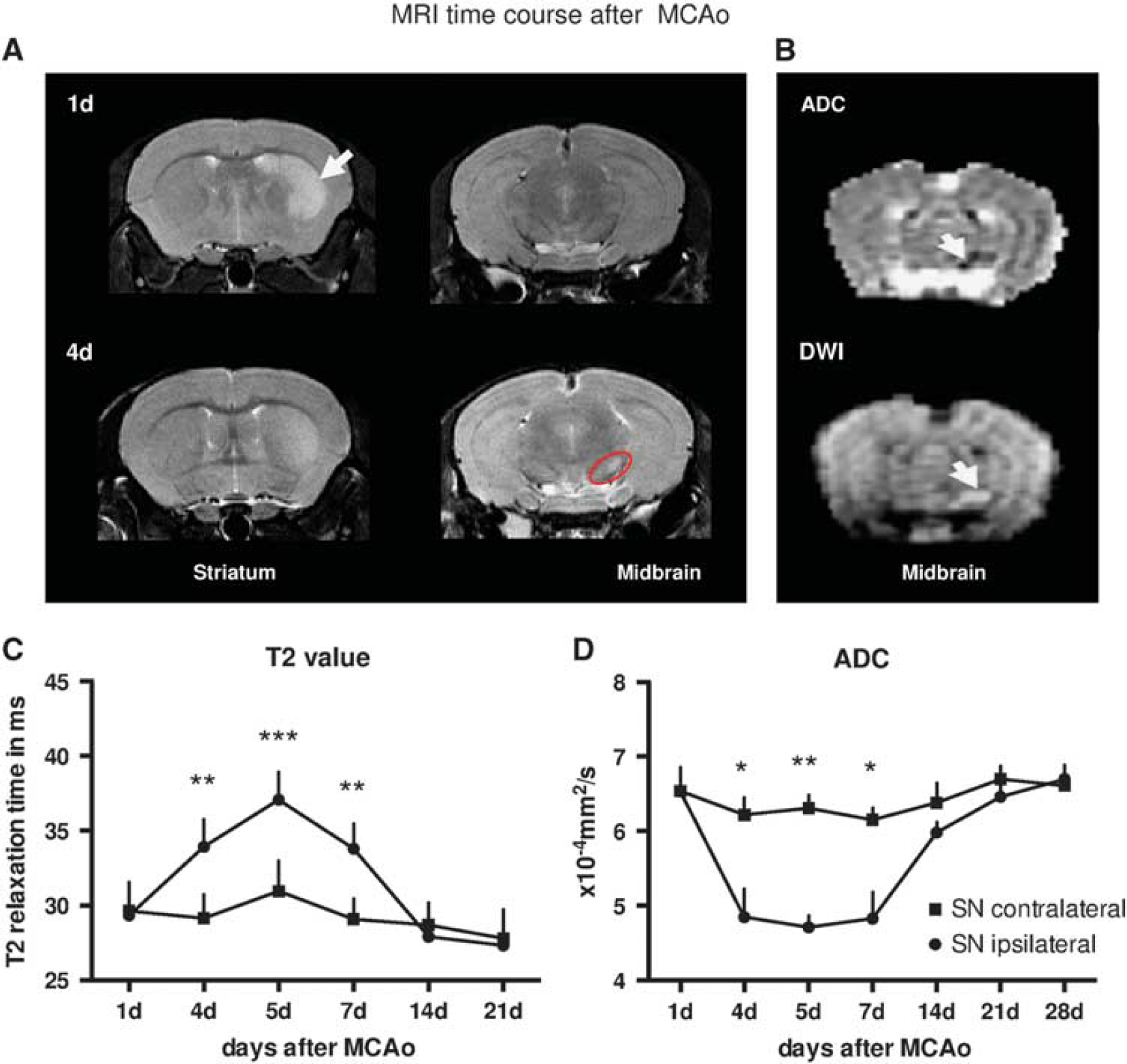
Magnetic resonance imaging (MRI) time course after middle cerebral artery (MCA) occlusion (MCAo). (
Immunocytochemical and Histologic Evaluation
Immunocytochemical and histologic evaluation of the ipsilateral SN as compared with the contralateral side showed no significant difference in the number of TH-immunoreactive and NeuN-positive cells on day 4 after MCAo, while Iba-1 cells showed a significant increase at this early stage. However, on day 7, the number of TH-positive as well as NeuN-positive cells had decreased significantly compared with the contralateral side. (Figures 2A–2C, 3A, and 3B). In both cell populations, NeuN- and TH-positive cells, the changes persisted until later time points, notwithstanding slight increases for NeuN-positive cells on day 28 compared with earlier time points (Figures 3C and 3D). Comparison across the different time points showed a highly significant decrease in the densities of ipsilateral NeuN-positive cells on day 7 relative to day 4 (
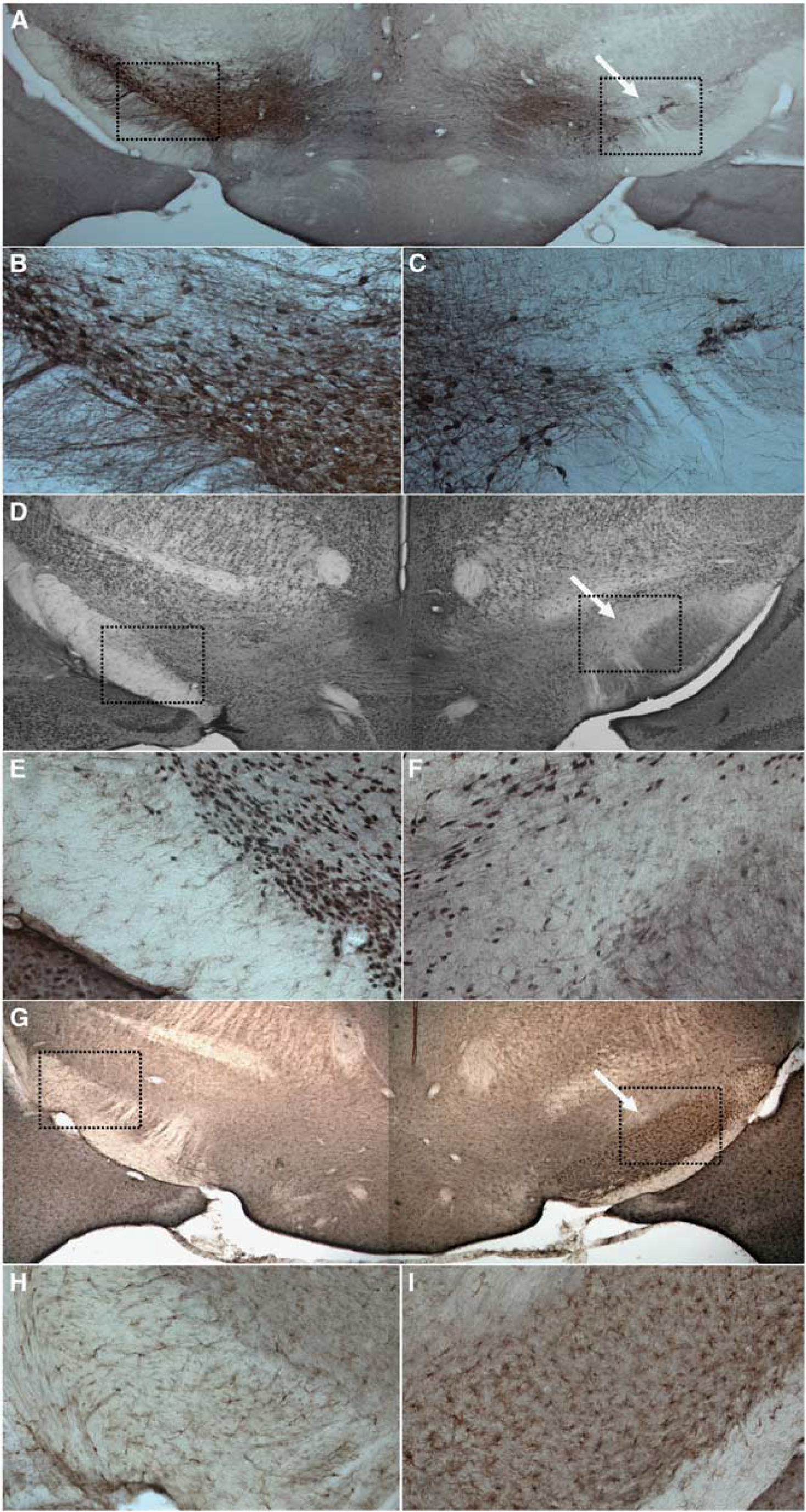
Nigral histology after middle cerebral artery occlusion (MCAo). Boxed areas in (
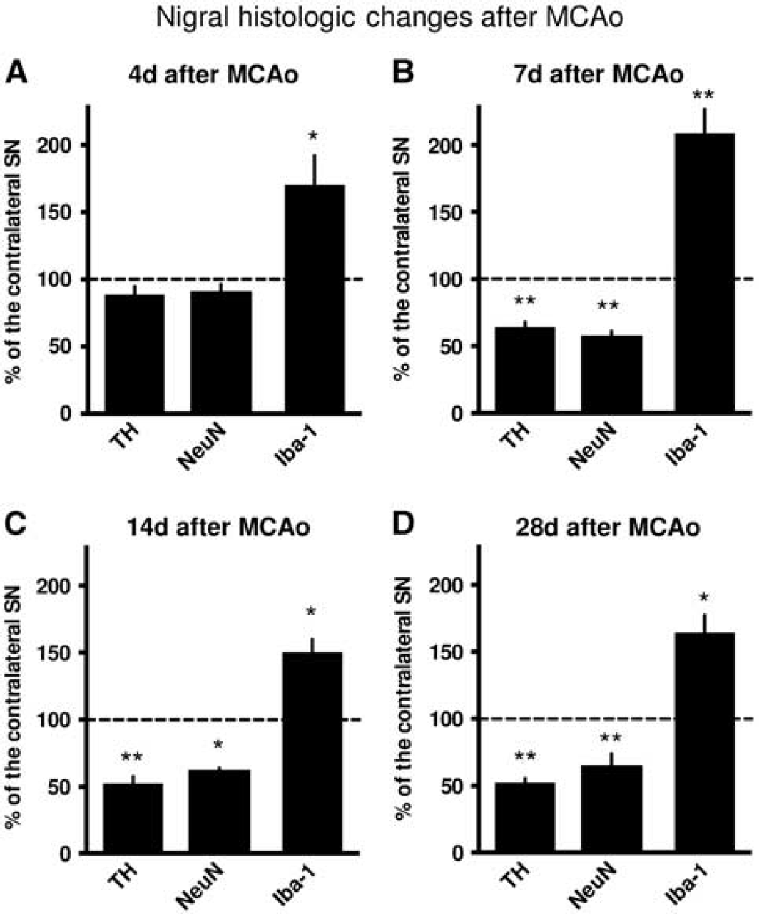
Nigral histologic changes after middle cerebral artery occlusion (MCAo). (
Treatment with FK506 and MK-801
In subsets of animals, treatment with MK-801 or FK506 was initiated on day 5 after 30 minutes MCAo/reperfusion. Treatment with MK-801, but not with FK506, conferred neuroprotection based on histologic evaluation on day 28 after MCAo (FK506,
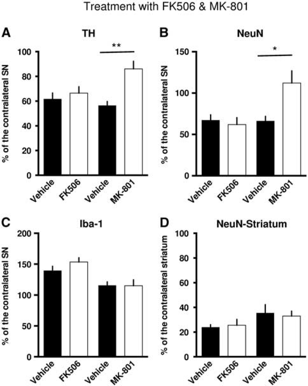
Treatment with FK506 and MK-801. (
DISCUSSION
This study has the following major findings: (1) Secondary neuronal degeneration in the SN occurs within a week of striatal ischemia and is characterized by marked neuronal cell loss. (2) Before significant cell loss on histology, transient MRI changes can be detected in the SN, indicating the onset of cellular edema and tissue damage. Our results suggest that MRI can be used to predict EPND in the SN after striatal infarction. (3) Treatment with MK-801, an NMDA receptor antagonist, but not with FK506, a potent anti-inflammatory drug, conferred neuroprotection even when treatment was initiated as late as on day 5 after MCAo. (4) Hence, our findings broaden the concept of neuroprotection after stroke and show a ‘delayed time window’ for potential interventions extending days after the ischemic event.
Together with the striatum, the SN forms a unique projection system that holds great importance for motor and behavioral functions. It has been suggested that EPND causes neurologic impairments that cannot be attributed to the primary ischemic lesion.6,9 If this were so, then EPND would be of great functional significance to stroke survivors. Furthermore, due to its delayed occurrence and prolonged development, EPND could also be a promising new target for neuroprotective treatments. The mechanisms underlying EPND are complex and only poorly understood. 7 So far, data from studies in mice are sparse. Our study therefore addressed the question of the dynamic changes involved in EPND in SN after striatal infarction. Furthermore, we investigated the effects of an anti-excitotoxic as well as of an anti-inflammatory drug on EPND when treatment was initiated in the subacute phase after MCAo.
Here, sequential neuroimaging examinations revealed transient MRI changes in the midbrain emerging as early as day 4 after MCAo. Significant alterations were apparent in ADC as well as in T2 values and clearly preceded histopathologic changes. Robust evidence of neuronal degeneration was only detectable from day 7 onwards. On day 7, a reduction in the density of NeuN-positive neurons to almost 50% of the contralateral side was found. This change persisted at later time points. In contrast, several studies in rats have suggested that the loss of TH immunoreactivity may only be transient.13,14 However, our data extending up to 28 days together with the results of Kronenberg
In line with nigral EPND, an increased number of activated microglia was found starting at 4 days after MCAo and thus before significant neuronal cell loss. Early inflammatory changes in the SN occurring before neuronal alterations have been reported recently.
8
Importantly, this finding might also explain the fact that, in our study, treatment with the calcineurin inhibitor FK506, a potent anti-inflammatory agent, failed to confer neuroprotection, since, in all probability, it was administered too late. However, treatment with MK-801 exerted neuroprotective effects even when treatment was begun on day 5 post event. This protective effect of MK-801 was borne out by separate analyses of NeuN+ and TH+ cells. The SN receives inhibitory GABAergic projections from the striatum and glutamatergic inputs from the subthalamic nucleus. The destruction of the GABAergic pathway after striatal ischemia may reduce inhibitory afferents to the SN, triggering increased neuronal firing with subsequently increased metabolism and finally neuronal cell death.
5
In line with this hypothesis, a protective effect of MK-801 against nigral degeneration has been shown previously, indicating the importance of transmission via NMDA receptors for EPND.
14
However, Yamada
Our study defines the dynamics of EPND in a murine stroke model, which is a crucial step to further elucidate the pathophysiologic mechanisms involved. These could prove to be valuable targets for the development of novel therapeutic approaches. The importance of characterizing new animal models for proper preclinical testing has just recently been emphasized. 22
CONCLUSIONS
This is the first study to establish and characterize a reliable model of EPND in the mouse using a multimodal approach with both neuroimaging and immunohistology. Secondary exofocal neuronal degeneration in the SN occurs within the first week of striatal ischemia and is characterized by marked neuronal loss, especially of TH+ dopaminergic neurons. In our study, transient MRI changes in the SN preceded overt signs of EPND on histology. Together with the striatum, the SN forms a unique dual projection system, which is of critical importance for behavioral and motor functions. It has been suggested that EPND may be responsible for neurologic deficits that cannot be attributed to the primary ischemic lesion. In particular, EPND of dopaminergic neurons in the midbrain (SN and ventral tegmental area) may contribute to motor impairments and affective sequelae after brain ischemia.6–9,23 Novel therapeutic approaches for the prevention of EPND after stroke may contribute to improved stroke outcomes.
Footnotes
VP and ME planned and designed the experiments. VP, AMH, GK, MB, and SM performed experiments. VP, AMH, CL, and GK analyzed the data and drafted the manuscript.
The authors declare no conflict of interest.