After the publication of this article, the following errors in the Materials and Methods section were noticed:
On page 1953 in the Materials and Methods section it is erroneously stated that ‘Cells were washed and incubated with rhodamine-conjugated, affinity-purified donkey anti-rat IgG (H+L) and fluorescein isothiocyanate (FITC)-conjugated, affinity-purified goat anti-rabbit IfG (H+L)’.
Correction: Cells were washed and incubated with rhodamine-conjugated, affinity-purified donkey anti-rabbit IgG (H+L) and fluorescein isothiocyanate (FITC)-conjugated, affinity-purified goat anti-mouse IgG (H+L).
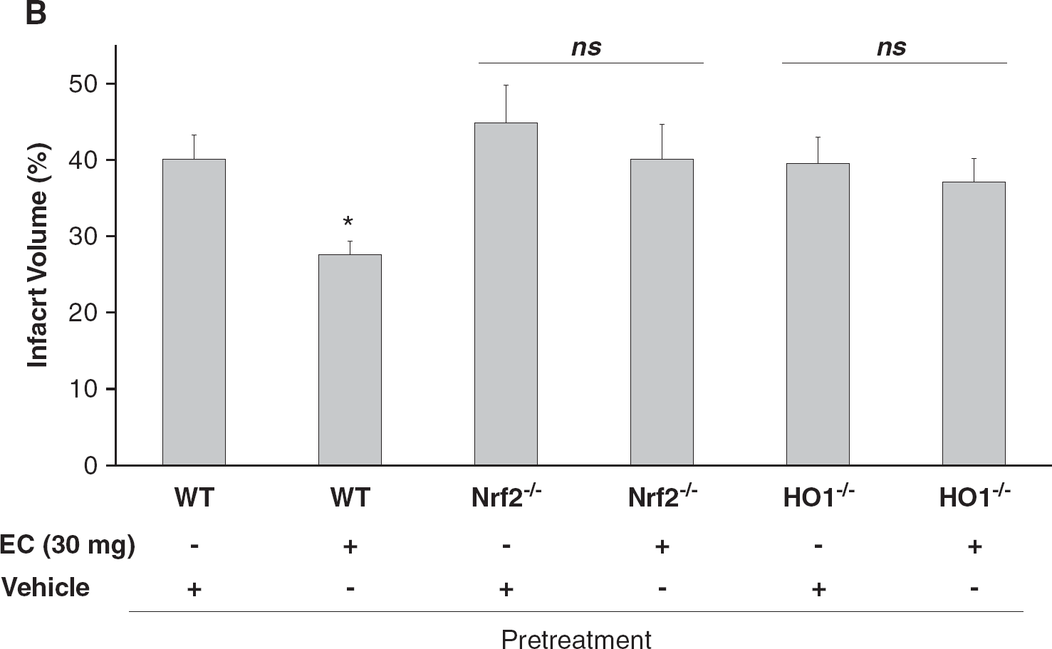
In Figure 5B the +and – representation is incorrect as published.
The corrected appears below.
The protective effect of epicatechin (EC) is lost in Nrf2−/− and HO1−/− mice. (A) A schematic diagram showing the pretreatment protocol of EC administration (90 minutes before middle cerebral artery occlusion (MCAO)) in Nrf2−/− and HO1−/− mice. (B, C) Nrf2−/− and HO1−/− mice and their wild-type (WT) littermates were treated with vehicle (n=10 WT, 7 HO1−/−, and 8 Nrf2−/−) or 30 mg/kg EC (n=11 WT, 7 HO1−/−, and 8 Nrf2−/−) 90 minutes before MCAO. At 24 hours after reperfusion, mice were tested for neurologic deficits and killed; their brains were collected and sectioned for TTC staining (note that the values for the WT mice are those from Figure 1 and are shown again here for comparison). (B) Quantification analysis revealed that the infarct volumes of EC-treated WT mice were significantly smaller than those of vehicle-treated WT mice. The infarct volumes of EC-treated Nrf2−/− and HO1−/− mice were not significantly different from those of their vehicle-treated counterparts, but were significantly higher than those of the EC-treated WT mice. (C) Similarly, the neurologic deficit scores of EC-treated Nrf2−/− and HO1−/− mice at 24 hours after MCAO were significantly higher than those of the EC-treated WT mice and not significantly different from those of their vehicle-treated counterparts. ∗P<0.05 compared with EC-treated WT controls; NS, not significant. (D) Neurons were grown for 24 hours in serum-free medium with B27 supplement minus antioxidant in the presence and absence of tert-butyl hydroperoxide (t-BuOOH; 60 μmol/L) or H2O2 (60 μmol/L), with or without EC (100 μmol/L), SnPPIX (10 μmol/L), or combinations thereof. Cell viability was assessed with the MTT assay. ∗∗∗P<0.001 versus vehicle; †P<0.05, ††P<0.01 versus t-BuOOH or H2O2; ‡‡‡P<0.001 versus t-BuOOH+EC or H2O2+EC. Values shown are the means ± s.e.m. of three batches of cells. (E) Primary postnatal cortical neurons isolated from HO1−/− or Nrf2−/− mice were incubated for 24 hours in Neurobasal medium alone (control) or in the presence or absence of t-BuOOH (60 μmol/L), EC (100 μmol/L), or combinations thereof. Cell viability was assessed with the MTT assay. ∗∗∗P<0.001 versus control; NS, not significantly different versus t-BuOOH.