
Abstract
Alzheimer's disease (AD) is a progressive, neurodegenerative disease that may involve inflammatory responses in the central nervous system (CNS). Our objective was to determine whether patients with amnestic mild cognitive impairment (aMCI), a preclinical stage of AD, have inflammatory characteristics similar to patients with multiple sclerosis (MS), a known CNS inflammatory disease. The frequency of lymphocytes and levels of pro-inflammatory cytokines in the cerebrospinal fluid of aMCI patients was comparable to MS patients or patients at high risk to develop MS. Thus, brain inflammation occurs early at the preclinical stage of AD and may have an important role in pathology.
INTRODUCTION
Previous studies of
MATERIALS AND METHODS
All subjects and/or their study partners signed the written informed consent approved by the Institutional Review Boards of the UT Southwestern Medical Center and Texas Health Presbyterian Hospital of Dallas, in accordance with the Federal-wide Assurance on file with the Department of Health and Human Services (USA).
Subjects
Eleven aMCI patients aged 57–76 years participated in the study. The diagnosis of aMCI was based on the Petersen criteria, 4 as modified by the Alzheimer's Disease Neuroimaging Initiative project (http://adni-info.org) using a multidisciplinary diagnostic conference format. Clinical evaluations were performed at the UT Southwestern Medical Center Alzheimer's Disease Center, to exclude other conditions that may cause memory problems. The mean score of the Mini-Mental State Examination was 29.3±0.8 and of the Wechsler Memory Scale Logical Memory for immediate and delayed recall were 11.0±1.4 and 9.4±0.9, respectively. All subjects had memory complaints and a global Clinical Dementia Rating score of 0.5 (memory box score 0.5). Subjects were screened to exclude clinical histories of stroke, major medical and psychiatric disorders, unstable heart diseases, uncontrolled hypertension, diabetes mellitus, and chronic inflammatory diseases.
For comparison, 23 CIS/MS patients aged 20–69 years who participated in the study were evaluated at the UT Southwestern Medical Center Neurology Clinic or associated hospitals for possible MS diagnosis. The CIS patients in this cohort (
CSF Collection
CSF collection from aMCI and CIS/MS patients was performed at the Department of Neurology and Neurotherapeutics at the University of Texas Southwestern Medical Center by lumbar puncture. The volume of CSF collected ranged from 4–12 mL.
Flow Cytometry
Flow cytometry was performed as previously described.
6
Briefly, the CSF was centrifuged for 10 minutes at 394
Enzyme-Linked Immunosorbent Assay for Cytokine and Antibody Output Methods
Methods for enzyme-linked immunosorbent assay for cytokine and antibody output are available in the Supplementary Information.
Statistics
All statistical analyses comparing aMCI and CIS/MS cohorts were performed using Student's
RESULTS
CSF cells stained with CD45 confirm their lymphocyte lineage (Figure 1A and 1B). aMCI patients averaged 66.8% CD45+ lymphocytes in the CSF cell population, whereas CIS/MS patients averaged 42.1% CD45+ lymphocytes. Interestingly, aMCI patients exhibited the similar wide range of CD45+ distribution as CIS/MS patients, in both lymphocyte percent (Figure 1A) and absolute number (Figure 1B), suggesting the presence of different degrees of brain inflammation in aMCI patients. These observations were also confirmed when only those CIS/MS patients of similar age to the aMCI patients were considered (data not shown). Notably, more than half of both the patient groups exceeded the absolute number of CD45+ lymphocytes typical of healthy donors 7 (660 lymphocytes/mL; data not graphed).
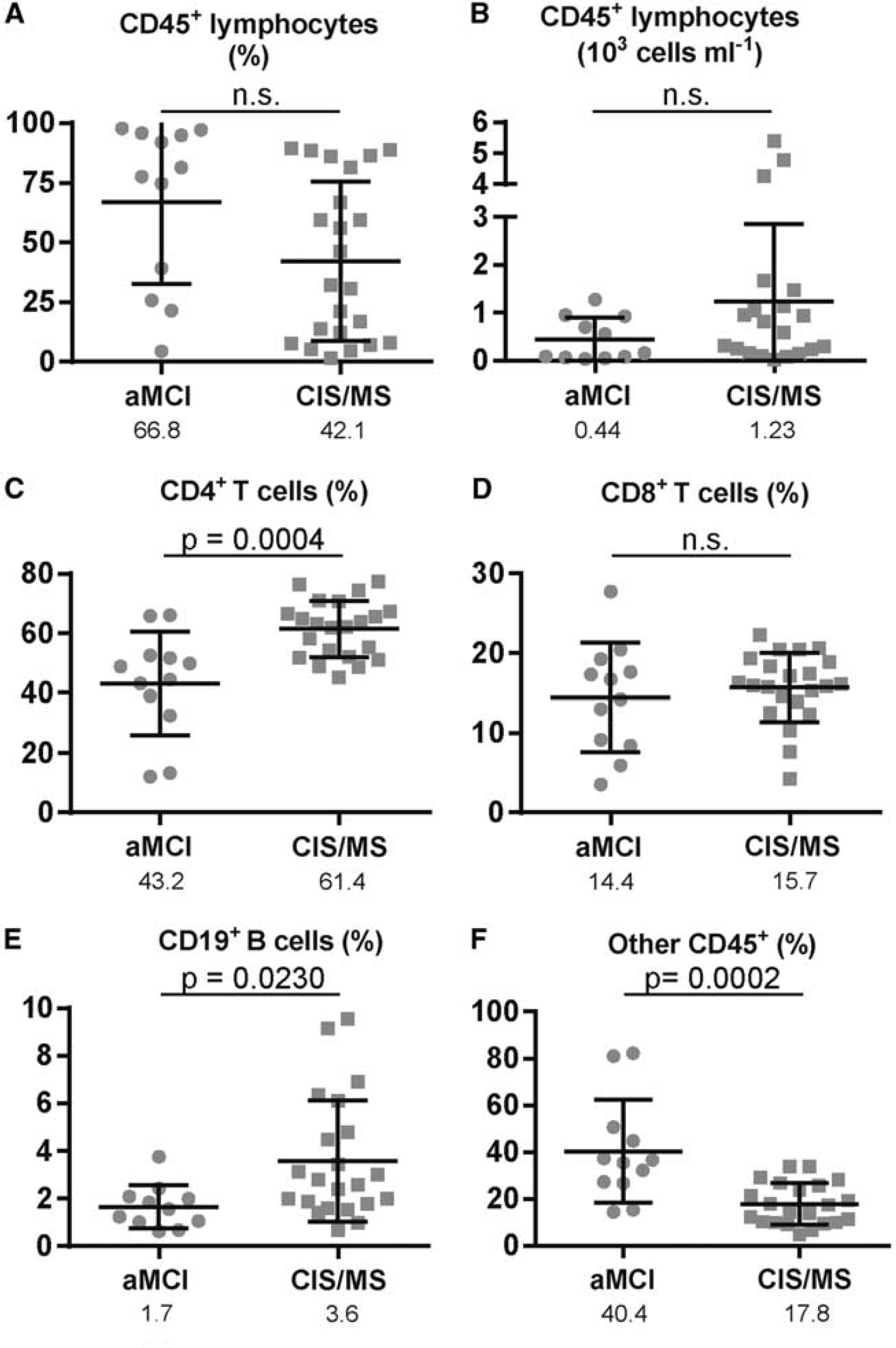
Lymphocyte dynamics in the cerebrospinal fluid (CSF) of amnestic mild cognitive impairment (aMCI) and clinically isolated syndrome/multiple sclerosis (CIS/MS) patients. CSF cells were pelleted and counted by a hemocytometer. (
CD4+ T cells are the predominant CSF lymphocyte population in CIS/MS patients (Figure 1C), in contrast to a reduced CD4+ T-cell population in aMCI patients (CIS/MS 61.4%; aMCI 43.2%,
Cytokines in the CSF that reveal MS-induced neuroinflammation include interferon-
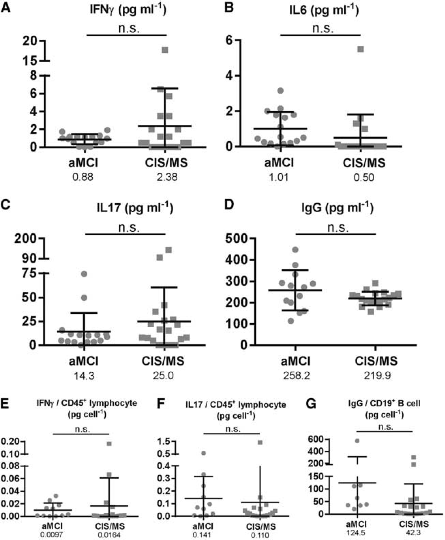
Cytokine and antibody profile in the cerebrospinal fluid (CSF) of amnestic mild cognitive impairment (aMCI) and clinically isolated syndrome/multiple sclerosis (CIS/MS) patients. CSF supernatants were evaluated for concentrations of interferon-
DISCUSSION
Patients with aMCI are at high risk to develop AD.
4
To our surprise, we found that CD45+ lymphocytes and pro-inflammatory cytokines were present in the CSF of aMCI patients at levels similar to those found in untreated patients with CIS or MS, the prototypical neuroinflammatory disease. To our knowledge, there are no previous studies of lymphocyte immune pathologic assessment
Early studies had identified CD4+ and CD8+ T cells in the
Elevated CSF lymphocyte counts correlate closely with the number of gadolinium-enhancing lesions, 12 and elevated CSF antibody production is one of the established indicators of MS diagnosis. 5 Recently, flow cytometry techniques have been used to characterize the immune cell profiles in the CSF of patients with MS13, 14 and CIS, who are at risk to develop MS. 15 These studies demonstrate that T cells, a critical component of the adaptive immune system, dominate the CSF of MS and CIS patients. B cells are also present at greater numbers in the CSF of MS and CIS patients compared with that in control cohorts. 14 In combination, these CSF lymphocyte profile studies included a total of 335 MS patients, 120 CIS patients, and 95 control patients. None of the previous cohorts included patients with AD or aMCI.
It is also intriguing that CSF from the aMCI patients contained levels of the pro-inflammatory cytokines IL6 and IL17 that were comparable to untreated CIS/MS patients. This suggests that inflammatory processes occurring in the brain of aMCI patients involve both cellular and cytokine components. We did not observe any correlation of IL6 or IL17 levels in the CSF with T-cell frequency in either the aMCI or CIS/MS cohort. Nevertheless, as IL6 and IL17 are central to the development of pro-inflammatory T cells,1, 8 it will be of particular importance to determine whether T cells from aMCI patients respond functionally to pro-inflammatory stimuli in a manner similar to that of T cells from patients who have classic neuroinflammatory diseases of the central nervous system (CNS), such as MS.
The findings of this study reveal that brain inflammation is likely to occur at the preclinical stage of AD, which can be assessed
Footnotes
The authors declare no conflict of interest.
ACKNOWLEDGMENTS
The authors thank the patients who participated in this study. We thank Charlene Supnet for editing of this manuscript. We thank C Hill and K Martin-Cook for recruiting and screening of study participants, cognitive data collection, and study coordination. We thank E Chang and P Chaudhury for CSF collection and processing. We thank MF Weiner, R Diaz-Arrastia, and BD Levine for clinical support.
References
Supplementary Material
Please find the following supplemental material available below.
For Open Access articles published under a Creative Commons License, all supplemental material carries the same license as the article it is associated with.
For non-Open Access articles published, all supplemental material carries a non-exclusive license, and permission requests for re-use of supplemental material or any part of supplemental material shall be sent directly to the copyright owner as specified in the copyright notice associated with the article.