
Abstract
Any tracer in fetal tissue comes from maternal arterial blood. Provided steady state is achieved and intermediate compartments are reversible, the Logan graphical methods should be applicable to the assessment of binding parameters in the fetal brain. Two pregnant rhesus macaques were studied with fallypride and the Logan method was used to assess dopamine receptor distribution volume ratios (DVRs) in both maternal and fetal striatum. The agreement between fetal striatal DVRs using maternal arterial blood and maternal and fetal cerebellum as input functions strongly supports our hypothesis that the conditions necessary for graphical analysis have been met.
Introduction
This work examines the application of conventional graphical analysis methods to the assessment of the brain dopaminergic system in the fetus of rhesus macaque using [18F]fallypride with maternal tissue or blood time courses as input functions.
Fallypride is a well-established dopamine D2/D3 receptor antagonist. Its high affinity for dopamine D2 receptor sites (in vitro KD = 30 pmol/L) (Mukherjee et al, 1995), selectivity, and rapid clearance from nonspecific binding sites make it a very favorable imaging agent for positron emission tomography (PET) brain studies. Fallypride PET tracer kinetics have been extensively modeled (Christian et al, 2000; Mukherjee et al, 2002), and its pharmacokinetics in nonhuman primate brains are well documented. For these reasons, [18F]fallypride was imaged in fetal nonhuman primate brains to show the utility of this tracer in the study of D2 receptor binding characteristics in the third trimester rhesus monkey fetus.
The fetus was treated as a maternal organ, as any exogenous compound introduced into fetal circulation comes from maternal arterial blood. Exchange of material between maternal and fetal blood occurs without actual mixing of the blood. We hypothesize that the assumptions required by the graphical approaches are met by the exchange of fallypride across the placenta between mother and fetus, allowing for meaningful estimations of distribution volumes (DVs) and distribution volume ratios (DVRs).
These studies show the utility of graphical analysis methods for assessing fetal dopaminergic receptor characteristics, specifically, the fetal striatal–cerebellar DVR.
Materials and methods
Radioligand Preparation
[18F]Fallypride was prepared according to published methods (Mukherjee et al, 1995) using the precursor (S)-N-[(1-allyl-2-pyrrolidinyl)methyl]-5-(3-tosyloxypropyl)-2,3-dimethoxybenzamide, purchased from ABX (Radeberg, Germany). Specific activities were on the order of 52GBq/μmol (1400 Ci/mmol).
Image Acquisition
Two healthy pregnant female rhesus monkeys (Macaca mulatta) (gestational days 127 and 128) were provided by the Wisconsin National Primate Research Center for use in these studies. The animal care and use procedures for all experiments were approved by the Institutional Animal Care and Use Committee of the University of Wisconsin Madison in compliance with NIH regulations. A dedicated Wisconsin National Primate Research Center anesthesiologist transported the animals, and maintained them on 1% to 2% isofluorane and monitored vital signs throughout the imaging procedure.
Animals were scanned on a GE Discovery LS PET/CT scanner, oriented perpendicularly to the length of the scanner bed in a left lateral decubitus position. This nontraditional orientation enabled dynamic imaging of the fetus and all maternal organs in a single bed position. Dynamic data were acquired in 2D mode in a sequence of eight 15 seconds frames, six 30-seconds frames, five 1-minute frames, five 2-minutes frames, four 5-minutes frames, and 11 10-minutes frames, initiated concurrently with bolus infusion of radiotracer. Venous blood samples of 900 μL were taken at 15, 30, 90, and 120 minutes after injection.
Images were reconstructed using an ordered subset expectation-maximization algorithm (2 iterations, 28 subsets) into a 256 × 256 × 35 image matrix with 1.95 × 1.95 × 4.25mm3 pixel size.
Plasma Analysis
Plasma metabolite analysis was based on previously published methods by Mukherjee et al (2002), who reported only small differences in the metabolite concentrations of venous versus arterial blood.
Positron Emission Tomography Data Analysis
All PET image analysis was done using the software package Amira 4.1 (Mercury Computer Systems, Chelmsford, MA, USA). Maternal whole blood activity concentration was determined by placing a region of interest (ROI) in the left ventricular chamber of the mother's heart. Lack of contamination from myocardial muscle accumulation of tracer into the image-based estimate of the maternal arterial time course was confirmed by comparison of multiple region volumes in the ventricular chamber.
The final 60 minutes of the data were averaged and used for determining the maternal and fetal brain ROIs by thresholding the PET data to exclude voxels below 50% of the maximum striatal value. Maternal ROI delineations were deemed reasonable, as the ROI volumes (2.31 to 2.56cm3) were consistent with a reported volume for male rhesus striatum (2.8 ± 0.08cm3) (Yin et al, 2009) adjusted for the 10% smaller total volume of the female rhesus brain relative to the male (Bourne, 1975; Yin et al, 2009). An elliptical ROI was placed on the cerebellar cortex, with the position verified by the corresponding computed tomography image. Fetal rhesus striatial ROIs had a volume of 0.75 cm3. The rhesus monkey fetal brain volume is approximately half that of the mother at 125 days gestation (Bourne, 1975), and thus this volume was slightly smaller than expected, probably because of partial volume effects.
The data were analyzed by the Logan arterial (Logan et al, 1990) and Logan reference region methods (Logan et al, 1996). The Logan arterial graphical analysis provides an estimate of the total DV of radioligand in the tissue of interest relative to plasma. The Logan reference method provides an estimate of the DVR, the total DV in a tissue of interest relative to the total DV in a reference tissue. A cerebellum reference region was chosen for its negligible dopamine receptor concentration. The well-established bias in the Logan slope estimates (Logan, 2000; Slifstein and Laruelle, 2000; Varga and Szabo, 2002) was controlled using the method of Varga and Szabo (2002). The Logan graphical analysis was implemented using IDL 6.3 (ITT Visual Information Solution, Boulder, CO, USA).
The maternal and fetal DVs for striatum and cerebellum were evaluated with the maternal blood as the input function. Ratios of these DVs provided the first of three estimates of the DVR of striatum and cerebellum in the fetal brain. The maternal and fetal striatal versus cerebellum DVRs were then directly evaluated with corresponding cerebellar time courses as the input functions. Finally, the maternal cerebellum was used as the input for a comparative DVR calculation of the fetal parameters (fetal striatum and fetal cerebellum). The ratio of these last two DVRs gave a third estimate of the DVR of the fetal striatum relative to fetal cerebellum as both of these DVRs were referred to the maternal cerebellar input function.
Results
Uptake and retention of [18F]fallypride was clearly seen in both the maternal and fetal striatum. Verification of fetal striatal accumulation was aided by the accompanying computed tomography image (Figure 1). Accumulation of ligand in fetal structures followed similar behavior as accumulation in maternal brain structures, although the fetal striatum had significantly lower accumulation of ligand compared with that in the mother.
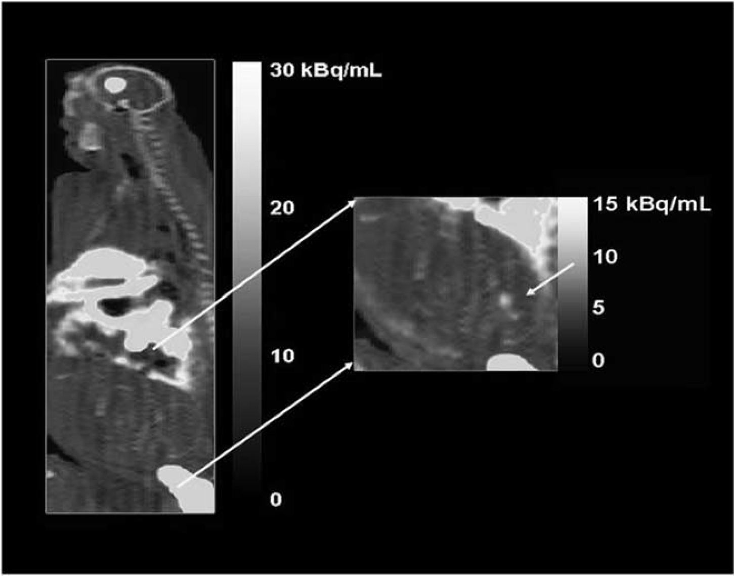
Positron emission tomography–computed tomography images show that both maternal and fetal regions can be assessed in a single bed position. Analysis of maternal brain and heart and fetal brain was possible from a single dynamic series. The uptake of fallypride in fetal striatum is clearly evident in the magnified fetal image (arrow).
Time–activity curves of the placenta and maternal arterial blood showed that over the last hour of the study, the placenta had the same relative rate of loss of concentration as the arterial blood. This supports our assumption of equilibrium between placenta and maternal blood, a condition necessary for graphical analysis.
Table 1 presents the DVR values for both mother and fetus using the various inputs. Fetal DVR values from all three approaches are seen to be consistent with each other.
DVR values for both maternal and fetal striatum evaluated using maternal arterial blood, maternal cerebellum, and fetal cerebellum
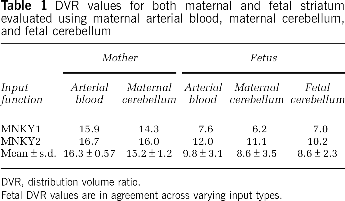
DVR, distribution volume ratio.
Fetal DVR values are in agreement across varying input types.
Discussion
Graphical analysis of reversible tracer kinetic data transforms the plasma and tissue uptake time courses into a linear plot, the slope of which is related to the number of available tracer binding sites. Graphical analysis is computationally easier and generally considered more robust for noisy data sets than compartmental analysis. Graphical analysis methods make no assumptions about compartmental model configurations, nor do they depend on the assumption that the reference fluid directly perfuses the tissue of interest. However, graphical analysis techniques do require several conditions to be met. The administered compound must be of tracer dose, all intermediate compartments must be reversible, and the system must attain steady-state equilibrium during the course of the study.
The results from this study show that the fetal DVR is measurable in utero noninvasively. The rhesus monkey fetus has a DVR of approximately half that of the mother in the third trimester of pregnancy. This is not directly comparable to human fetal brain development, as the size of a rhesus monkey brain at birth is ∼60% to 70% that of its adult size, whereas a human brain is only about 25% of its adult size at birth (Gibson, 1991; Stauber et al, 1974).
The placental barrier may cause some concern with regard to these data sets, because of the possibly complex nature of exchange. It is proposed here that the exchange of materials across the placenta has no bearing on the DVR of the fetal striatum once steady state is achieved. Once all tissues suffer the same fractional loss of concentration versus time, the intermediate components along the path from maternal arterial plasma to fetal brain will equilibrate with each other. In other words, the placenta has an equilibrium DV relative to maternal arterial blood and the umbilical vein has an equilibrium DV relative to placenta. In this chain of intervening reversible components connecting maternal plasma to fetal brain, the concentration in the numerator of each DVappears as the denominator in the next. If all intervening tissues have truly reached equilibrium, the graphical estimates of the DV in the fetal brain are as valid as if the brain were perfused by maternal blood directly. The fact that the intermediate compartments involve several organ systems instead of all being in the brain does not prevent this situation from conforming exactly to the stipulations originally set by Logan et al (1990, 1996).
If the placenta or some intermediate organ were to irreversibly bind fallypride, or if steady state were not reached, then the conditions of the graphical methods would not have been achieved and the fetal DVRs calculated using three different inputs would not agree. Irreversible metabolism of the tracer in the fetus would also violate the steady-state assumption. The agreement among the DVR values further implies that any additional metabolism by the fetus was slow enough to maintain steady-state conditions to a good approximation.
Compartmental analysis of these data was not possible without a known compartmental configuration. Use of even a simple compartmental configuration would require significant sampling from fetus, placenta, and uterine cavity to assess the behavior of these compartments, making the studies extremely invasive. In addition, identifying fetal heart or umbilical vein from the PET data as the inputs for fetal tissue was not a possibility as each structure represents a combination of both arterial and venous blood signal.
Conclusion
This study shows that the uptake and kinetics of [18F]fallypride can be simultaneously measured noninvasively in both maternal and fetal brains. In addition, graphical methods currently used for calculating DVRs using fallypride are capable of identifying those same properties in a third trimester fetus. The input from either the mother's arterial blood or cerebellum yield the same results as using the fetal cerebellum. Therefore, it is possible to study fetal dopaminergic development noninvasively. Further assessment of the behavior of tracers within the fetus using compartmental models requires significant invasive sampling from mother, placenta, uterine cavity, and fetus. Provided all the necessary conditions are met, graphical methods yield the desired results.
Footnotes
The authors declare no conflict of interest.