
Abstract
Traumatic brain injury (TBI) frequently results in neuroinflammation, which includes the invasion of neutrophils. After TBI, neutrophils infiltrate the choroid plexus (CP), a site of the blood—cerebrospinal fluid (CSF) barrier (BCSFB), and accumulate in the CSF space near the injury, from where these inflammatory cells may migrate to brain parenchyma. We have hypothesized that the CP functions as an entry point for neutrophils to invade the injured brain. Using the controlled cortical impact model of TBI in rats and an
Keywords
Introduction
The choroid plexus (CP) is located in all four cerebral ventricles. It is a highly vascularized tissue, in which blood microvessels are enclosed by a single layer of cuboidal epithelial cells (Strazielle and Ghersi-Egea, 2000). The CP is the major source of cerebrospinal fluid (CSF); however, together with the arachnoid membrane, it also constitutes the blood—CSF barrier (BCSFB). Increasing evidence indicates that the BCSFB has an important role in brain immune response and neuroinflammation (Strazielle and Ghersi-Egea, 2000). Choroidal venules have been shown to express P- and E-selectins, and it has been proposed that the BCSFB is involved in the recruitment of T cells to the CSF (Kivisäkk et al, 2003). We, along with other researchers, have shown that, after traumatic brain injury (TBI), neutrophils infiltrate the lateral ventricle CP and accumulate in the cistern of velum interpositum, a part of the third ventricle, and in the subarachnoid CSF space near the injury site, from where these inflammatory cells appear to migrate to traumatized brain parenchyma (Carlos et al, 1997; Chodobski et al, 2003). These findings suggested that not only the blood—brain barrier but also the BCSFB and CSF pathways are involved in neutrophil invasion observed after TBI. Neutrophil invasion has been shown to contribute to the formation of posttraumatic brain edema (Schoettle et al, 1990), and treatments directed to counter neutrophil influx to the injured brain showed several beneficial effects (Matsumoto et al, 1997; Yamasaki et al, 1997; Beech et al, 2001).
The accumulation of neutrophils in the CP occurring after injury indicated that the choroidal tissue has the ability to produce neutrophil chemoattractants; however, such properties of the CP have not yet been investigated. Neutrophil chemoattractants belong to the family of CXC chemokines (Kobayashi, 2008). Cytokine-induced neutrophil chemoattractant (CINC)-1 (referred to as CXCL1), CINC-2α and CINC-2β (both referred to as CXCL3), and CINC-3 (also known as macrophage inflammatory protein-2 or MIP-2; referred to as CXCL2) are the four major rat chemokines that possess neutrophil chemotactic activity (Huang et al, 1992a,
b
; Nakagawa et al, 1994). Various types of epithelial cells have previously been shown to synthesize CXC chemokines on exposure to proinflammatory cytokines, such as tumor necrosis factor-α (TNF-α) and interleukin-1β (IL-1β) (Takaishi et al, 2000; Handa et al, 2004). It has also been shown that both basolateral and apical secretion of chemokines is necessary for promoting the transepithelial migration of neutrophils (McCormick et al, 1995, 1998). The aim of this study was to define a role of the CP in neutrophil recruitment to the injured brain. Accordingly, we characterized the ability of the CP to produce CXC chemokines in response to injury using a rat model of TBI and determined the direction of cytokine-dependent release of these chemokines (across the apical versus the basolateral membrane of the choroidal epithelium) using an
Materials and methods
Reagents and Antibodies
ThermoScript RNase H− reverse transcriptase and RNase inhibitor, RNaseOut, were obtained from Invitrogen (Carlsbad, CA, USA). HotStart
The Rat Traumatic Brain Injury Model
Adult male Long-Evans rats weighing 250 to 350 g (Harlan, Indianapolis, IN, USA) were used for gene expression analysis, whereas adult male Sprague-Dawley rats (Charles River, MA, USA) of the same weight were used for other studies. They were kept at 22°C on a 12-h light cycle and maintained on standard pelleted rat chow and water
Real-Time Reverse-Transcriptase Polymerase Chain Reaction
At 1, 2, 4, and 6 h, and 1, 2, and 4 days after TBI, rats (9 to 10 animals per time point) were reanesthetized with pentobarbital sodium. Samples of the lateral ventricle CP, both ipsilateral and contralateral to injury, were collected separately and pooled into three subgroups (three to four rats per subgroup). Samples of the cerebral cortex adjacent to the lesion and those of the contralateral cortex from the corresponding region were collected. Samples of the lateral ventricle CP and those of the cerebral cortex were also collected from sham-injured rats. Total RNA was isolated using the NucleoSpin RNA II kit (Macherey-Nagel, Düren, Germany). First-strand cDNAs were synthesized using oligo(dT)20 primer (50 pmol) and 15 U of ThermoScript RNase H− reverse transcriptase. Forty units of RNase inhibitor, RNaseOut, were also added to the reverse transcription reactions. For each reaction, 0.5 μg of total RNA was used and the reactions were performed for 1 h at 50°C.
Real-time PCR was performed using the TaqMan chemistry in a DNA Engine Opticon System (MJ Research, Waltham, MA, USA). The sequences of primers and TaqMan probes, and the predicted sizes of PCR products are given in Table 1. Cyclophilin A was used for the normalization of obtained data. The 50-μL PCR reaction mixtures contained 0.2 mmol/L of mixed dNTPs (deoxyri-bonucleotide triphosphates), 0.2 μmol/L of each primer, 0.1 μmol/L of TaqMan probe, 5 mmol/L of MgCl2, 1 U of HotStart
The sequences of primers and TaqMan probes, and the predicted sizes of PCR products
CINC-3, cytokine-induced neutrophil chemoattractant-3; IL-1β, interleukin-1β; MIP-2, macrophage inflammatory protein-2; PCR, polymerase chain reaction; TNF-α, tumor necrosis factor-α.
F, forward primer; R, reverse primer; R probe.
Immunohistochemistry
Rats (two to four animals per group) were reanesthetized with pentobarbital sodium and were perfused transcardially with ice-cold 0.9% NaCl, followed by ice-cold 4% paraformaldehyde in 0.05 mol/L of phosphate-buffered saline (PBS), pH 7.4. Brains were removed and postfixed for an additional 4 h in the paraformaldehyde—PBS solution at 4°C. They were then incubated overnight in 20% sucrose in PBS and embedded in the Tissue-Tek OCT compound (Sakura Finetek, Torrance, CA, USA). Coronal brain sections were cut on a cryostat at 10 μm.
Immunohistochemical procedures were performed at room temperature, except for the incubation with primary antibodies that was completed at 4°C. All incubations were performed in PBS containing 0.25% BSA and 0.25% Triton X-100. For washes, PBS containing 0.1% BSA and 0.1% Triton X-100 was used. To minimize nonspecific staining, the brain sections were incubated for 30 mins with 10% normal goat serum (Jackson Immunoresearch Labs, West Grove, PA, USA). Overall 4% of the normal goat serum was also added when the sections were incubated with primary and secondary antibodies. After the initial blocking step, the brain sections were incubated overnight with primary antibodies. The following concentrations of primary antibodies were used: 1 μg/mL for CINC-1 and IL-1β, 5 μg/mL for TNF-α, 13.2 μg/mL for MPO, 0.4 mg/mL for Golgi 58 K protein, 2 μg/mL for CD11b, 10 μg/mL for NKCC1, and 2.5 μg/mL for β-catenin. Six 10-min washes were then performed, and the sections were incubated for 1 h with secondary antibodies at a concentration of 2 μg/mL. After four 10-min washes, the brain sections were mounted with a Vectashield mounting medium (Vector Labs, Burlingame, CA, USA).
Myeloperoxidase Activity Assay
To quantitatively assess the magnitude of posttraumatic accumulation of neutrophils in the choroidal tissue, the MPO activity assays were performed. Rats (four animals per group) were reanesthetized with pentobarbital sodium and were perfused transcardially with ice-cold 0.9% NaCl. Samples of the lateral ventricle CP, both ipsilateral and contralateral to injury, those of the cerebral cortex adjacent to the lesion, and those of the contralateral cortex from the corresponding region were collected. They were homogenized at room temperature in 50 mmol/L of PPB (potassium phosphate buffer), pH 6.0, and centrifuged for 20 mins at 60,000 ×
Transmission Electron Microscopy Analysis
At 1 day after TBI or sham injury, rats (two to six animals per group) were reanesthetized with pentobarbital sodium, and the samples of the lateral ventricle CP, both ipsilateral and contralateral to injury, were removed and fixed overnight in 2.5% glutaraldehyde in 0.15 mol/L of sodium cacodylate buffer, pH 7.4, at 4°C. After fixation, the specimens were treated with 1% OsO4 in cacodylate buffer for 1 h at 4°C. They were then dehydrated in a graded acetone series and embedded in Epox 812 resin (Fullam, Latham, NY, USA). Ultrathin sections (50 to 60 nm) were prepared, retrieved onto 300 mesh copper grids, and contrasted using uranyl acetate and lead citrate. Sections were examined using a Morgagni 268 transmission electron microscope (FEI, Hillsboro, OR, USA).
Primary Cell Cultures of Rat Choroidal Epithelial Cells
The procedures were conducted according to the guidelines approved by the French Ethical Committee (decree 87 to 848) and by the European Community directive 86-609-EEC. Animals were obtained from Harlan (Gannat, France). Choroidal epithelial cells were isolated as previously described (Strazielle and Ghersi-Egea, 1999), and were seeded on Transwell Clear inserts (Costar, Cambridge, MA, USA) with the pore size of 0.4 μm precoated with laminin. The cells were kept at 37°C in a humidified atmosphere of 5% CO2/95% air and were fed every other day with a medium containing 10% fetal bovine serum. The experiments were carried out 5 days after the cells reached confluence.
Secretion of Cytokine-Induced Neutrophil Chemoattractant-1
The cells were serum starved in a serum-free medium supplemented with 0.1% BSA for 24 h before experimentation. On the day of the experiment, the BSA-containing serum-free medium was renewed in both chambers, and cytokine treatment was initiated by exposing the apical surface of the cell monolayers to various concentrations of rat IL-1β for different periods of time. In time-course studies, the cells were incubated with 0.1 ng/mL of IL-1β for 1.5, 4, 8, and 24 h. In dose-response studies, the cells were exposed to 0.01, 0.1, 1, and 10 ng/mL of IL-1β for 8 h. Control monolayers (incubated in the serum-free medium without IL-lβ) were processed simultaneously. At the end of the incubation periods, media were collected from the apical and basolateral chambers and stored at −80°C until further processing for CINC-1 quantification. The epithelial monolayers were subsequently analyzed for possible changes in paracellular permeability. The concentration of CINC-1 in the culture media was determined using ELISA (enzyme-linked immunosorbent assay) using the Quantikine Rat CINC-1 Immunoassay kit from R&D Systems Europe. The samples were assayed in duplicates after appropriate dilution.
Basolateral-to-Apical Permeability to Cytokine-Induced Neutrophil Chemoattractant-1
Cells were serum starved as described above, and their basolateral surface was exposed to either recombinant CINC-1 (0.4 or 4 ng/mL) or to conditioned media collected from basolateral chambers in another set of epithelial cell monolayers treated for 4 h with 0.1 ng/mL of IL-1β. After 1.5 and 4 h of incubation, media were sampled from both compartments to assess the CINC-1 concentration. No degradation of recombinant or endogenous (conditioned media) CINC-1 during the incubation periods was observed.
Measurement of Paracellular Permeability of Epithelial Monolayers
The integrity of epithelial monolayers was examined by measuring the permeability of each filter to the paracellular permeability marker, 14C-sucrose. The permeability × surface area product, PSt, was calculated from the 14C-sucrose clearance rates as previously described (Strazielle and Preston, 2003). The permeability coefficient, Pt, was calculated by dividing PSt by the surface area of the cell monolayer.
Statistical Analysis
For statistical evaluation of data, ANOVA (analysis of variance) was used, followed by the multiple comparison tests, as described in figure legends.
Results
Posttraumatic Increase in mRNA for Proinflammatory Cytokines and CXC Chemokines in the Choroidal Tissue and the Injured Cortex
The CP is not only a target for proinflammatory cytokines, such as TNF-α and IL-1β (Ericsson et al, 1995; Nadeau and Rivest, 1999), but is also a site of their synthesis (Quan et al, 1999). Accordingly, we first examined posttraumatic changes in choroidal expression of TNF-α and IL-1β using real-time reverse-transcriptase -PCR. Trauma resulted in triphasic changes in TNF-α expression in the ipsilateral CP. Compared with sham-injured rats, there was a rapid (within 1 h after TBI) increase in TNF-α mRNA, followed by a gradual decrease and a secondary increase at 6 h after TBI (Figure 1A). Interestingly, we also observed an increase in TNF-α expression in the contralateral CP at 1 h after TBI, but not at later time points after injury. The levels of TNF-α expression in the injured cortex were slightly higher than those found in the CP (Figure 1C). The time course of posttraumatic changes in TNF-α mRNA was similar to that found in the CP, except that the secondary increase in mRNA levels at 6 h after TBI was not observed. No changes in TNF-α expression in the contralateral cortex were observed. The time course of changes in choroidal IL-1β expression differed from that found for TNF-α. The message for IL-1β in the ipsilateral CP increased gradually after injury to reach a peak at 6 h after TBI (Figure 1B). The synthesis of IL-1β in the contralateral CP was also upregulated at 4 and 6 h after TBI compared with sham injury; however, the levels of mRNA for IL-1β found in the contralateral CP were significantly lower than those observed in the ipsilateral tissue. The levels of IL-1β mRNA in the injured cortex were higher than those found in the ipsilateral CP (Figure 1D); however, no changes in IL-1β expression in the contralateral cortex were observed.
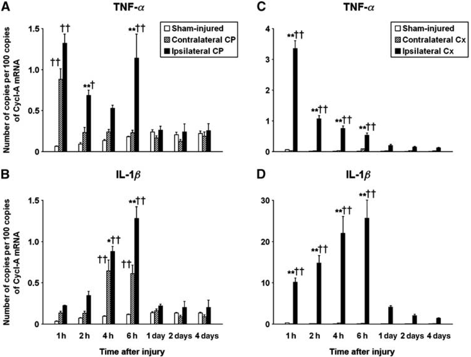
Real-time RT-PCR analysis of temporal changes in choroidal (CP) and cortical (Cx) expressions of TNF-α and IL-1β after TBI. The controlled cortical impact model of TBI in rats was used. (
Posttraumatic changes in choroidal expression of three neutrophil chemoattractants, CINC-1, CINC-2α, and CINC-3/MIP-2, were examined. The expression levels of CINC-2β were found to be low (data not shown). The maximum increase in mRNA for all three chemokines in the ipsilateral CP was observed at 6 h after TBI, with the levels of chemokine synthesis being 120- to 430-fold higher compared with those found in sham-injured animals (Figures 2A–2C). The expression of CINC-2α in the contralateral CP was also upregulated at 4 and 6 h after TBI and that of CINC-3/MIP-2 tended to increase during the same period of time after injury; however, the levels of mRNA for these chemokines found in the contralateral CP at 6 h after TBI were significantly lower than those observed in the ipsilateral CP. By contrast, the induction of CINC-1 synthesis was limited to the ipsilateral CP. The peak in expressions of CINC-1 and other CXC chemokines (observed at 6 h after TBI) was followed by an abrupt decrease at 1 day after trauma, and the levels of chemokine expression remained low until the end of the 4-day observation period. The levels and the time course of changes in expression of these chemokines in the injured cortex were similar to those found in the CP, but the upregulation of chemokine synthesis in the contralateral cortex was not observed at any time point after TBI (Figures 2D–2F).
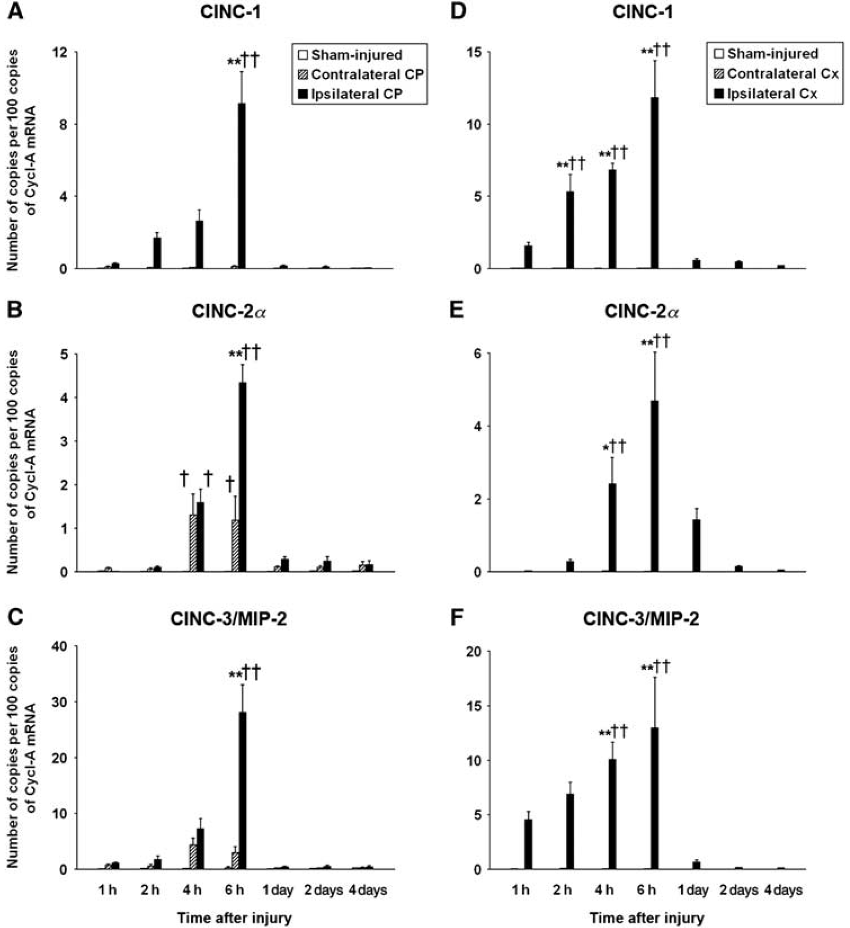
Real-time RT-PCR analysis of posttraumatic changes in choroidal (CP) and cortical (Cx) expressions of neutrophil chemoattractants. (
Choroidal Expression of Proinflammatory Cytokines and Cytokine-Induced Neutrophil Chemoattractant-1 at the Protein Level
The immunohistochemical analysis of the choroidal tissue harvested at 8 h after TBI showed the TNF-α-positive staining of epithelial cells in the ipsilateral CP (Figure 3A). Colocalization of this staining with the immunostaining for the NKCC1 cotransporter, an integral membrane protein expressed apically in the choroidal epithelium (Praetorius and Nielsen, 2006), suggested that this TNF-α-immunoreactive product represents the transmembrane precursor form of TNF-α (Kriegler et al, 1988). No TNF-α-immunopositive staining was observed in the contralateral CP (data not shown). In the ipsilateral CP, IL-1β was highly expressed in epiplexus (Kolmer) cells (Ling et al, 1998), but not in stromal macrophages (Figure 3B). In the contralateral CP, epiplexus cells were negative for IL-1β (data not shown). Interestingly, IL-1β also appeared to be constitutively expressed in the choroidal epithelium (Figure 3C). The IL-1β-immunoreactive product was associated with the Golgi complex, as shown by double immunostaining with monoclonal antibody to the Golgi marker, Golgi 58 K (Bloom and Brashear, 1989), and was found in both the ipsilateral and contralateral CPs, as well as in the CP from sham-injured rats (data not shown). Intense cytoplasmic CINC-1-positive staining of epithelial cells in the ipsilateral, but not in the contralateral CP was also associated with the Golgi complex (Figure 3D). The localization of CINC-1 to the Golgi complex is in line with increased synthesis of this secreted protein occurring after TBI. In addition, the CINC-1-immunopositive staining was localized to the basolateral membrane of choroidal epithelial cells (Figure 3E), and this pattern of staining was similar to that found for ion transporters expressed basolaterally in the choroidal epithelium (Praetorius and Nielsen, 2006). The basolateral CINC-1-positive staining did not colocalize with the staining for β-catenin, a component of the adherens junction complex, indicating that CINC-1 is confined to a part of the basolateral membrane that is distinct from areas occupied by adherens junctions (Figures 3E and 3H). As chemokines have the ability to bind to heparan sulfates (Kuschert et al, 1999), it is possible that the basolateral localization of CINC-1 was associated with the binding of this secreted protein to heparan sulfate proteoglycans expressed on the basolateral surface of the choroidal epithelium (Fayein et al, 1992; Snow et al, 1994). The levels of CINC-1 expression varied in the choroidal tissue with some epithelial cells showing strong CINC-1-immunopositive staining and others bearing weaker CINC-1 immunoreactivity. These variable levels of CINC-1 expression appeared to result in an uneven distribution of neutrophils infiltrating the choroidal tissue observed after injury (data not shown). Double immunostaining for CINC-1 and NKCCl showed that the CINC-1-immunoreactive product is not associated with the apical membrane domain (data not shown). In the contralateral CP, only a weak basolateral staining for CINC-1 was observed (Figure 3F). This pattern of staining was similar to that found in the choroidal tissue collected from sham-injured animals (data not shown).
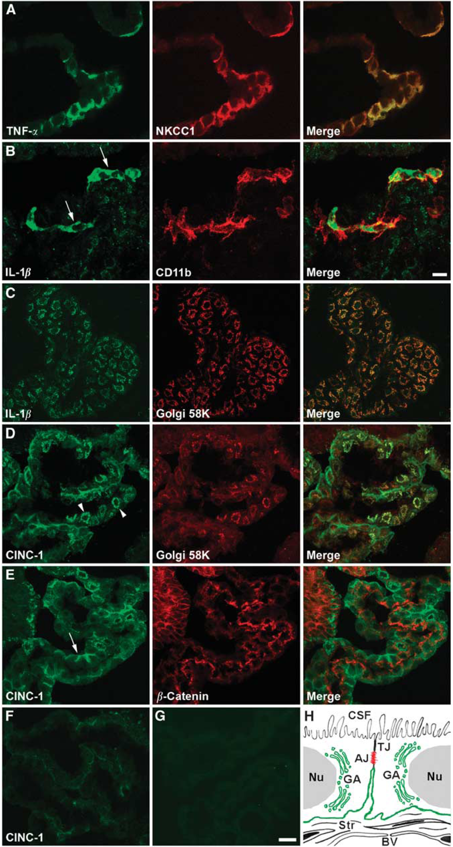
Immunohistochemical localization of tomor necrosis factor-α (TNF-α), interleukin (IL)-1β, and cytokine-induced neutrophil chemoattractant (CINC)-1 in the lateral ventricle CR Confocal microscopy images of the choroidal tissue harvested at 8 h after TBI are shown. (
Polarity of Cytokine-Induced Neutrophil Chemoattractant-1 Secretion by the Choroidal Epithelium
To test whether IL-1β induces CINC-1 synthesis in the choroidal epithelium and to determine the direction of CINC-1 release (across the apical versus the basolateral membrane), we used an
Under control conditions, CINC-1 was constitutively produced, but it was barely detectable, and the total amount of CINC-1 secreted to the apical and basolateral chambers during the 24-h observation period was ~7 pg per filter. This secretion was polarized, with CINC-1 being largely released across the apical membrane. In time-course studies, the cells were incubated with 0.1 ng/mL of IL-1β for 1.5, 4, 8, and 24 h (Figure 4A). At this concentration of IL-1β, CINC-1 secretion was predominantly basolateral throughout the 24-h observation period. During the initial 4-h period of exposure to IL-1β, the CINC-1 levels in the apical and basolateral chambers increased rapidly. However, between 4 and 8 h of incubation with this cytokine, the CINC-1 levels increased at a much slower rate, and the concentrations of CINC-1 in both the apical and basolateral chambers at 24 h of exposure to IL-1β were not significantly different from the respective CINC-1 levels found at the 8-h time point. In dose-response studies, the cells were exposed to 0.01, 0.1, 1, and 10 ng/mL of IL-1β for 8 h (Figure 4B). A 32-fold increase in CINC-1 secretion over its constitutive production was induced by IL-1β at 10 pg/mL. Similar to control conditions, CINC-1 was largely released across the apical membrane of the choroidal epithelium. The exposure of epithelial monolayers to IL-1β at 100 pg/mL resulted in a ~600-fold increase in CINC-1 production and the polarity of CINC-1 secretion was reversed, with approximately two-third of the chemokine produced by choroidal cells being secreted across the basolateral membrane. The production of CINC-1 reached a plateau when IL-1β was applied to epithelial monolayers at 1 ng/mL.
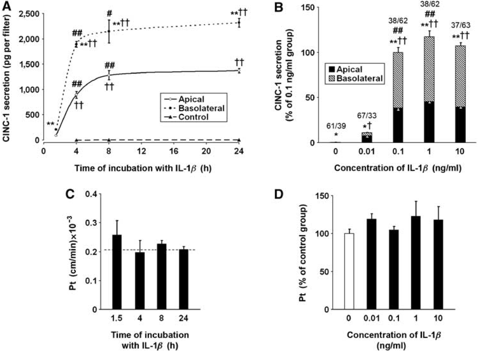
Secretion of cytokine-induced neutrophil chemoattractant (CINC)-1 in primary cell cultures of rat choroidal epithelial cells. The cells were seeded on Transwell Clear inserts as described in the section, Materials and methods. The experiments were performed 5 days after the cells reached confluence (
The possible effect of the leak and/or transcellular transfer of CXC chemokines on our estimates of polarity of CINC-1 secretion by epithelial monolayers was evaluated. To this end, the basolateral surface of the monolayers was exposed to conditioned media collected from IL-1β-treated cells. The monolayers were exposed for 1.5 h to the conditioned medium containing 513 pg/mL of CINC-1 and for 4 h when the medium contained 2.62 ng/mL of CINC-1. The levels of CINC-1 measured in the apical chamber at the end of the incubation periods were higher than those resulting from the constitutive secretion, which was assessed in parallel. However, the amounts of CINC-1 that actually permeated the monolayers represented only 0.53 ± 0.13 to 0.57 ± 0.10% of the amount of the chemokine present in the basolateral chamber. Similar low basolateral-to-apical permeability to CINC-1 was observed when the recombinant chemokine was applied at 0.4 and 4 ng/mL for 1.5 and 4 h, respectively (data not shown). The exposure of epithelial monolayers to IL-1β or CINC-1 did not cause any apparent alterations in the integrity of the BCSFB, as shown by the lack of changes in paracellular permeability to sucrose (Figures 4C and 4D, data not shown).
Neutrophil Trafficking Across the Blood—Cerebrospinal Fluid Barrier
Posttraumatic induction of choroidal synthesis of chemokines was followed by neutrophil recruitment to the ipsilateral CP; however, there was a significant delay between an increase in chemokine production and neutrophil accumulation in the choroidal tissue (Figures 5A–5F). Neutrophils infiltrating the ipsilateral CP were found at 1 day after injury, whereas at 8 h after TBI, these inflammatory cells were only sporadically observed to accumulate in the ipsilateral CP. This neutrophil influx was transient, as no neutrophils were found to infiltrate the ipsilateral CP at 2 days after TBI. Neutrophils did not accumulate in the contralateral CP (Figures 5A, 5C, and 5E), which was consistent with the absence of posttraumatic induction of CINC-1 synthesis in the contralateral CP, and only a limited upregulation of expression of CINC-2α and CINC-3/MIP-2 occurring in this tissue (see Figures 2A–2C). These immunohistochemical data were confirmed by the MPO activity assays. Low, but consistently detectable, MPO activity was found in the contralateral CP. It is likely that stromal macrophages and/or epiplexus cells contributed to this low MPO activity (Nagra et al, 1997). At 8 h after TBI, there was a slight, but significant, increase in MPO activity in the ipsilateral CP when compared with the contralateral CP (Figure 5G). Consistent with immunohistochemical findings, MPO activity in the ipsilateral CP peaked at 1 day after injury. Interestingly, increased MPO activity in the ipsilateral CP was also found at 2 days after injury, which was likely associated with the accumulation of monocytes observed at that time point after TBI (unpublished observations). A similar pattern of changes in MPO activity was found in the cerebral cortex adjacent to the lesion, but the peak levels of MPO activity (at 1 day after TBI) were approximately five-fold higher than those observed in the ipsilateral CP (Figure 5H). Myeloperoxidase activity was undetectable in the contralateral cortex.
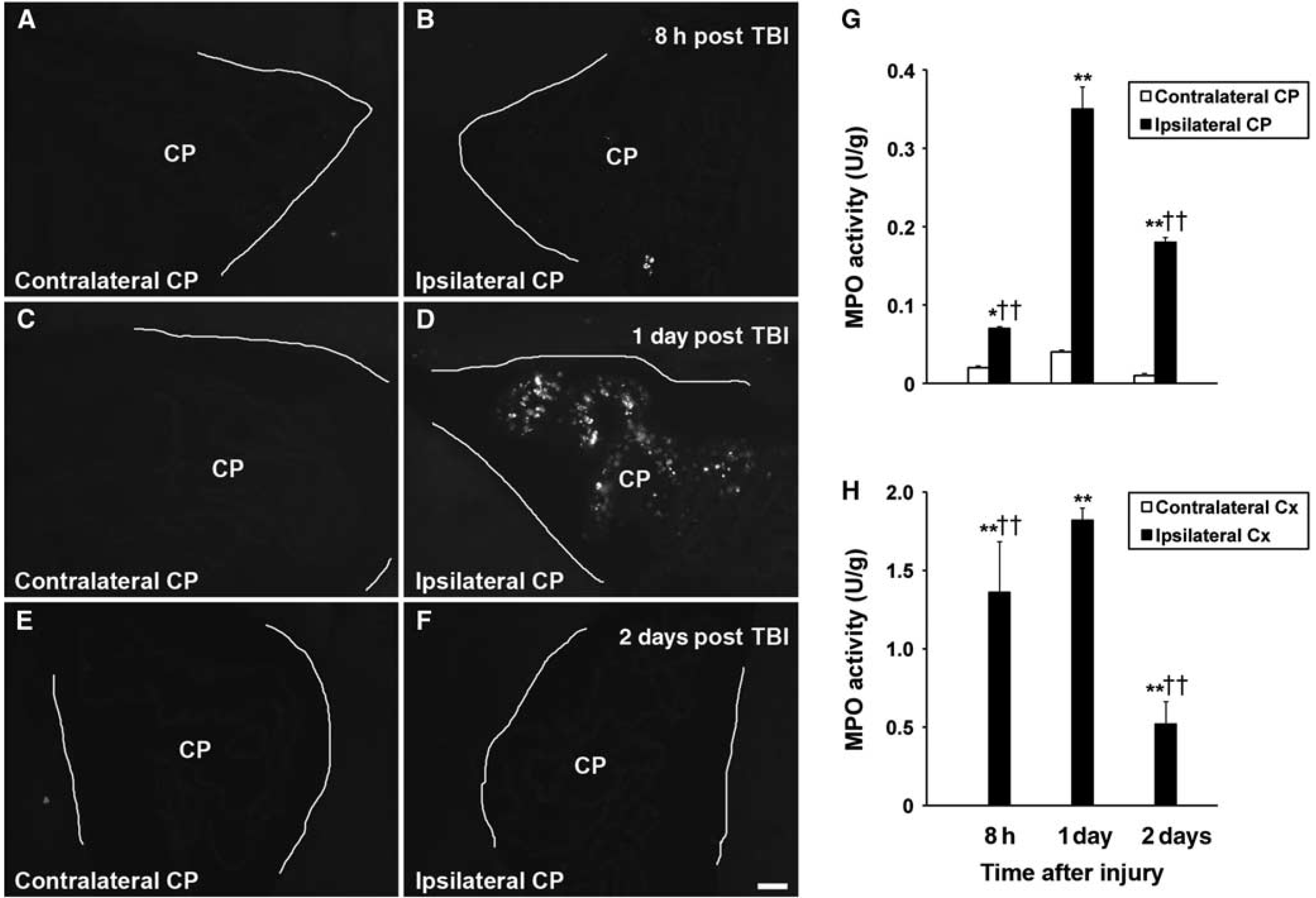
Posttraumatic accumulation of neutrophils in the choroid plexus (CP). (
To show neutrophil trafficking across the BCSFB, we used transmission electron microscopy. The contralateral CP harvested at 1 day after TBI had normal morphology (Figure 6A), similar to the morphology of the choroidal tissue from sham-injured rats (data not shown). Choroidal epithelial cells had well-defined microvilli and the lateral membrane was folded into elaborate interdigitating processes near the base of the cells. The space between adjacent epithelial cells was narrow and the tight junctions between the cells were well discernible. In the ipsilateral CP, neutrophils were found to infiltrate the stroma and subsequently reach the intercellular space between epithelial cells (Figures 6B and 6C). The movement of neutrophils between epithelial cells toward their apical domain did not appear to affect the integrity of tight junctions.
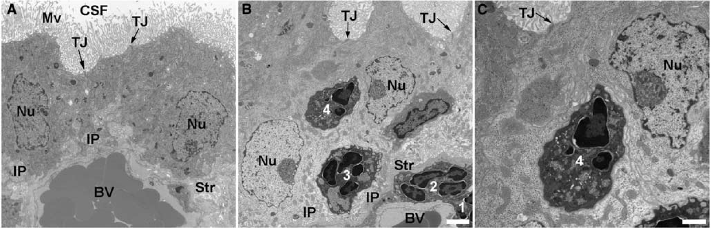
Transmission electron microscopy (TEM) analysis of the CR The choroidal tissue was harvested at 1 day after TBI and processed for TEM. (
Discussion
Brain injury is associated with a rapid and substantial increase in the synthesis of proinflammatory cytokines, such as TNF-α and IL-1β, in traumatized brain parenchyma (Fan et al, 1995, 1996). In this study, we found that, after TBI, the expression of TNF-α and IL-1β is also upregulated in the CP located ipsilaterally to the injury. Therefore, an increase in CSF levels of proinflammatory cytokines observed in TBI patients (Singhal et al, 2002; Buttram et al, 2007) may result from the production of these inflammatory mediators by both brain parenchymal cells and the choroidal tissue. Proinflammatory cytokines are strong inducers of epithelial synthesis of CXC chemokines (Takaishi et al, 2000; Handa et al, 2004), and we observed a gradual increase in choroidal expression of these chemokines after TBI. It is likely that CSF-borne proinflammatory mediators, including TNF-α and IL-1β, have a major role in inducing the production of neutrophil chemoattractants in the CP, and that this choroidal chemokine production is, in part, augmented by the CP-derived cytokines. Neutrophils themselves are also able to produce CXC chemokines and can respond with increased chemokine synthesis to inflammatory stimuli, such as lipopolysaccharide (Scapini et al, 2000). However, the posttraumatic increase in expression of CXC chemokines observed in the ipsilateral CP was not related to neutrophil accumulation in the choroidal tissue. In fact, there was a considerable delay between an increase in the choroidal synthesis of chemokines and neutrophil invasion of the CP, and at 1 day after TBI, when neutrophils infiltrated this tissue, the mRNA levels in the ipsilateral CP for all three chemokines studied were close to those found in the contralateral CP or in the CP from sham-injured rats. Interestingly, an increase in chemokine expression in both the ipsilateral CP and in the ipsilateral cerebral cortex peaked at approximately the same time after injury (6 h after TBI), but the time courses of neutrophil influx to these brain regions differed. Indeed, neutrophil invasion to the injured cortex was occasionally observed as early as 4 h after TBI and was consistently observed at 6 to 8 h after injury (Chodobski et al, 2003), whereas in the CP, these inflammatory cells were only observed sporadically at 8 h after TBI. These observations suggest that factors other than chemokine synthesis, such as e.g., different time courses of induction of expression of cell adhesion molecules on choroidal and cerebrovascular endothelium, account for different kinetics of neutrophil migration to the CP and traumatized brain parenchyma.
Specific targeting by neutrophils of the ipsilateral CP located at some distance from the site of injury is unique. The recruitment of inflammatory cells to the CP has previously been observed after intraparenchymal injection of IL-1β, and it occurred in the absence of leukocyte influx to the site of injection (Andersson et al, 1992). It is likely that the volume-transmission-mediated signaling involving bulk flow of interstitial fluid has a critical role in choroidal induction of chemokine synthesis and in the resultant recruitment of neutrophils (Redzic et al, 2005). Volume transmission may also have a role in posttraumatic increase in expression of proinflammatory mediators observed in the contralateral CP and in the contralateral hippocampus (data not shown). Consistent with this idea, unilateral injury to the cerebral cortex has been found to promote the bulk flow of interstitial fluid along the white matter fibers of corpus callosum from the ipsilateral to the contralateral hemisphere (Kamada et al, 1995). We have also observed an increase in chemokine synthesis in the fourth ventricle CP, although the levels of expression of these inflammatory mediators in this tissue were lower than those found in the lateral ventricle CP ipsilateral to injury (data not shown). As CSF flows from the lateral ventricles to the third ventricle and thereafter continues its movement along the cerebral aqueduct and the fourth ventricle, the induction of chemokine synthesis in the fourth ventricle CP was likely mediated by volume transmission involving the bulk flow of CSF (Redzic et al, 2005). Despite increased chemokine synthesis, in neither the fourth ventricle CP nor the CP from the lateral ventricle contralateral to injury was neutrophil recruitment observed. It is possible that, in these tissues, the levels of expression of CXC chemokines were too low and/or there was insufficient induction of expression of cell adhesion molecules on choroidal microvasculature for promoting neutrophil migration.
To characterize the inducibility and direction of chemokine release from the choroidal epithelium, we used an
The above-discussed features of the choroidal epithelium, such as the ability to synthesize CXC chemokines in response to injury and bidirectional secretion of these chemokines across the apical and basolateral membranes of choroidal epithelial cells, strongly suggest that the BCSFB has a role in posttraumatic invasion of neutrophils. This idea is further supported by our transmission electron microscopy analysis of the choroidal tissue. Similar to the movement across the blood—brain barrier (Wong et al, 2007), the migration of neutrophils across the BCSFB was found to involve the paracellular pathway and did not appear to affect the integrity of tight junctions.
Footnotes
Acknowledgements
We thank Ms Julie Sarri for her technical assistance, Ms Virginia Hovanesian for her help in acquiring and processing confocal microscopy images, and Ms Carol Ayala for her help in electron microscopy procedures.
The authors declare no conflict of interest.