
Abstract
Protein analysis (using either specific protein quantitation by methods such as HPLC and immunoassays or structural analysis by methods such as LC-MS) usually requires significant sample preparation, including quantitative purification of the target protein from complex sample matrices and potentially enzymatic treatment or labeling. We have developed platform for high-throughput microchromatography, capable of running 96 or more small volume samples in parallel, producing from 10 pg to 100 μg of purified protein from each sample.
The platform is based on disposable cartridge devices with 5 μL packed bed of resin. The cartridges may be operated as spin columns or run on a modified 96-channel liquid handler with ultra-low dead volume syringes that directly connect to the cartridges, providing very precisely controlled positive-displacement flow control. A major application is quantitative purification of target proteins using affinity or physical chromatography. Using large diameter nonporous beads, standard microplate enzyme-linked immunosorbent assay reagents can be used to perform 30-min immunoassays. Enzymatic digestion methods have also been developed on the system for application in glycan profiling.
Keywords

Introduction
The growing importance of biotherapeutics to the pharmaceutical industry has created an ever-increasing demand for high-precision, high-sensitivity, and high-throughput protein analysis. Protein analysis is used throughout the development process, starting with research protein expression systems during the discovery phase, changing to serum samples during lead optimization, characterization, preclinical and clinical trials (both for the biotherapeutic itself and related protein biomarkers), and ending with cell culture supernatants during the scaling up and operation of the commercial manufacturing process. Complex samples to be analyzed range from research protein expression systems to biological samples such as serum to cell lysates and cell culture supernatants used for commercial production.
There are two general objectives for protein analysis. One is to accurately and precisely quantify the amount of a specific protein present in the sample. Several techniques are widely used for specific protein quantitation, including HPLC and immunoassays. All of these methods involve using either physical methods or (more commonly) affinity methods to selectively separate the target protein from the complex sample, followed by a sensitive detection method (usually based on optical absorbance, fluorescence, chemiluminescence, or mass spectrometry).
The other objective of protein analysis is to determine the overall structure, particular structural features, and/or bioactivity of the target protein. A wide range of analytical instrumentation (such as LC-MS and nuclear magnetic resonance) is used for structural analyses of proteins to examine characteristics such as the peptide sequence or posttranslational modifications. In most cases, however, the target protein must first be selectively purified intact from the complex sample matrix. Many structural analysis methods also involve other complex preparation methods (such as enzymatic treatment or labeling) before instrumental analysis.
Both specific protein quantitation and structural analysis usually require complex multistep workflows to prepare samples for final instrumental readout. Most of these workflows involve combinations of liquid handling for dilution and reagent addition, highly selective separations, and enzymatic treatment. In this article, we report on a new platform for high-throughput microliter-scale protein chromatography and enzymatic treatment, which enables a very wide range of complex protein analysis sample preparation workflows to be carried out on standard microplate-based liquid-handling automation systems.
Technical Requirements
Over the last 50 years, liquid chromatography has proven to be a highly robust and versatile general technique for protein purification from complex samples, both for analytical and preparative applications. The challenge has been to adapt liquid chromatography to the requirements of high-throughput analysis. To meet this challenge, a number of critical technical requirements must be met.
Scale
Analytical samples in biopharmaceutical development are often quite small in both volume and mass of target protein present, are often quite precious, and in many cases are needed for several different purposes. Sample volumes are often in the range of 10s to 100s of microliters, with target protein mass in the range of picograms (or even femtograms) to micrograms. Typical preparative chromatography systems are usually designed for samples that are orders of magnitude larger.
Throughput
The throughput requirements for protein analysis vary widely, depending on the application. However, a common theme across the biopharmaceutical industry is that throughput requirements are increasing, driving a need to consider new technical approaches. For many applications, batches of several hundred to several thousand samples at a time are not uncommon. Traditional liquid chromatography is relatively low throughput, as systems are designed to run one sample at a time in serial fashion. Even with modern Ultra-HPLC, runs require at least several minutes from injection to injection. More widely available preparative media or enzyme reactions can require much longer run times. To reach the throughputs required for many applications, significant parallel processing is clearly required.
Quantitative Binding and Elution
If chromatography is to be used to quantitate the target protein across a range of sample concentrations and matrices, it is critical that the binding, washing, and elution of the analytical target are nearly complete and highly reproducible. Even if the application only involves qualitative structural analysis, quantitative binding and elution can still be highly desirable to prevent biasing of the results between variant forms of the protein.
A key requirement for complete binding is sufficient exposure time of the sample to the resin to both allow the target protein to reach the binding surface (mass transport) and interact with the selective binding groups on the surface (binding kinetics). The former is often limiting because of the slow diffusion rates of large protein molecules, 1 but the latter can also be limiting in some cases, particularly in the case of some antibody/antigen interactions. 2
Efficient washing away of nonbinding impurities and complete elution of the target also require sufficient time for mass transport, although the need is usually much less stringent than for quantitative binding. However, it is also important to completely separate the free washed or eluted molecules from the resin. This is very difficult to do quantitatively when the resin is used in a batch or equilibrium adsorption mode in which a volume of the wash or elution buffer is allowed to come to equilibrium with a volume of resin. Multiple steps are always required, resulting in a fairly large dilution factor. If a packed bed is used in the more normal flow-through mode, quantitative washing and elution can be accomplished in as few as five-bed volumes in a single step.
Control of Flow Rate
One key to obtaining quantitative binding and elution is to control the flow rate through the chromatography column. For example, with agarose resins frequently used for protein purification, residence times of around 5 min are required for the dynamic binding capacity to approach the equilibrium capacity. 3 With a 5-μL packed bed of resin, this translates to a flow rate of only 1 μL/min. Even with more modern “high-speed” resins, residence times of 0.5–1 min are needed, necessitating flow rates of 5–10 μL/min. Achieving these controlled flow rates across an entire 96-well plate at once is a major challenge.
Air Entrainment
Trapping of air bubbles in a chromatography bed is a problem even for conventional methods, but the effects are greatly magnified as the scale is reduced. With a 5-μL packed bed, even a miniscule 1-μL air bubble can cause a major loss of capacity or recovery. The system used must be both designed to minimize air entrainment in routine operation and deal with it reliably if it occurs.
Format
The Society for Biological Screening/American National Standards Institute microplate format has become the standard for handling protein analysis samples, and any high-throughput microchromatography system should be fully compatible with this standard. However, because of the wide variation in the number of samples to be processed in different applications, systems with 96 columns in a single unit are less desirable.
Previous Approaches
A number of previous approaches have been tried for high-throughput microchromatography of proteins, but each has suffered from significant drawbacks. One approach has been to form a packed resin bed in the distal end of a pipette tip. A number of methods have been used for doing this, and pipette tip columns with a number of common protein resins are available, with bed volumes ranging upward from the 5- to 10-μL range. Air displacement is used to aspirate and dispense liquid in and out of the tip and thus through the bed. Because of this, the flow rate is both highly variable and uncontrolled, and air entrainment is difficult to prevent. In addition, because there is only one entry and exit port through the packed bed, washing and elution operate in batch equilibrium mode, necessitating multiple steps and high dilution factors. Although useful for qualitative extraction, pipette tip columns have never been successful for quantitative chromatography.
Another common approach is to use packed bed cartridges in conjunction with a vacuum manifold to drive liquids through the bed using air pressure. This method is used very frequently and successfully for solid-phase extraction of small molecules and can often provide fully quantitative results. However, because of the much lower diffusion rates for proteins, the residence times required for quantitative binding are much higher than for small molecules, and it is difficult or impossible to turn down the vacuum enough to achieve the low, controlled flow rates needed. In addition, vacuum manifold systems often suffer from foaming with high-protein content samples.
Cartridge Technology
The AssayMAP high-throughput microchromatography system (BioSystem Development, LLC, Madison, WI) was designed specifically to meet the technical requirements of quantitative sample preparation for protein analysis. The AssayMAP system consists of a microchromatography cartridge together with some special laboratory ware. The cartridge and laboratory ware system is designed to support two modes of operation—spin format (using the cartridges as spin columns, with centrifugal force driving the liquid through the bed) and probe syringe format (using the cartridges with a special liquid handler to provide precise flow control). In both cases, the system is adapted to work with conventional microplate laboratory automation hardware.
The AssayMAP cartridge (Fig. 1) 4 is designed around a 5-μL bed volume packed with resin. The bed is contained between two polymeric support filters that are molded into the cartridge housing. Any resin with a particle size range of roughly 15–150 μm can be used in the cartridge, including polymeric, agarose, or silica-based materials. Resins have been used in virtually all of the common chromatographic modes and can also be used with immobilized enzymes. The resin is packed into the bed under highly controlled conditions and slight compression to insure stability in both flow directions and reasonably consistent hydraulic permeability.
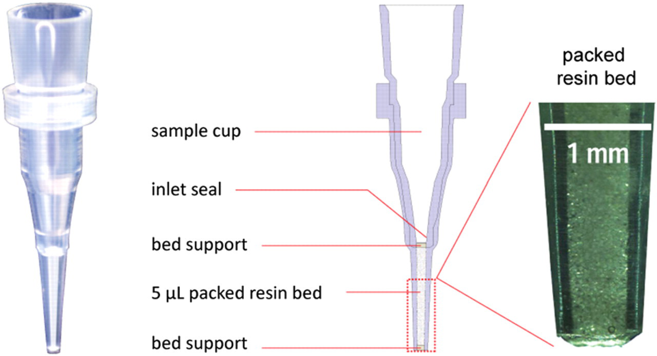
AssayMAP cartridge, showing major components and a photomicrograph of the outlet tip, with the packed bed and insert-molded bed support filter.
The resin bed is contained within a housing that has approximately the same outlet tip geometry as a pipette tip to enable small volumes of liquid to be precisely aspirated and dispensed through the bed from a variety of common sample containers, including 384-well microplates. Immediately above the upper bed support is a small conical inlet seal, which can function similarly to a miniature tapered Luer-type fitting to connect the bed directly to the liquid handler as described below. Above the inlet seal is a sample cup with a volume capacity of 200 μL, which is used when the cartridge is operated as a spin column. The inside of the sample cup incorporates some small ribs that enable the cartridge to be picked up by standard laboratory pipettes but prevent an airtight seal from being formed so that liquid cannot be accidentally displaced through the bed by air pressure (which can cause air entrainment).
AssayMAP cartridges are supplied in a molded rack (Fig. 2 A) that holds 96 units in an 8 × 12 array and mounts on virtually any standard 96-well microplate, with the cartridges held precisely centered with their outlet tips just below the entrances of the wells. The cartridges fit loosely in the rack so that the liquid-handling system can pick them up easily without disturbing the rack. The rack is also designed so that the liquid-handling head can pick up fewer than 96 cartridges if desired.
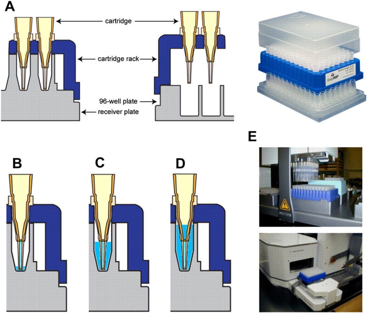
(A) Cartridge laboratory ware system, including rack and two types of collection plates—special receiver plates and standard 96-well microplates. (B) Spin format operation with small volumes using the receiver plate. As little as 5 μL through the bed causes the cartridge outlet tip to be immersed in liquid, preventing drying. (C) Up to 120 μL may be completely passed through the cartridge. (D) With 200 μL loaded, the bed is completely immersed in liquid after centrifuging to equilibrium, enabling use of high-speed (1000 ×g) spins to remove entrapped air. (E) The spin format may be automated using conventional liquid- and plate-handling robotics.
The cartridges are also supplied with a special molded “receiver plate” that fits below the rack. The cartridges hang into the wells of the receiver plate with their tips just above the well bottom, and the top of the packed bed is below the inlet of the well. The receiver plate thus protects the cartridges and can keep the resin bed fully immersed during shipping if required. During operation on an automated system, the receiver plate provides support to the rack to keep it from flexing, ensuring reliable pickup of cartridges. The receiver plate also performs a number of critical functions when the cartridges are operated in the spin format.
Spin Format
In one mode of operation, the cartridges function as spin columns in which the cartridges, racks, and collection plates (normal 96-well microplates or the special receiver plate) are run in a microplate centrifuge to drive liquid through the packed bed. The flow rate in this case is controlled by the g-force (revolutions per minute) of the centrifuge, together with the hydraulic permeability of the cartridge, viscosity of the liquid, and height of the liquid pressure head across the packed bed. The system has been engineered to meet the minimum residence time requirements for quantitative binding and elution in a single pass for typical samples using standard equipment and reagents. Resins with relatively small particle sizes (typically 20–50 μm) and enhanced rapid mass transport properties are generally required for good performance in the spin format.
This method uses low g-forces (50 ×g for sample loading and 200–300 ×g for washing and elution) to generate the required residence times. The design of the receiver plate/rack/cartridge system minimizes the risk of air entrainment in the packed bed or air bubbles just above the bed, which can lead to a stopped flow (air locking). The 5-μL packed bed only contains 2–4 μL of liquid; so drying can also lead to air locking. As shown in Figure 2B, even very small volumes of liquid (∼5 μL) passed through the bed into the receiver plate well are sufficient to immerse the cartridge tip and keep the bottom of the bed from drying out. Surface tension and capillary action (wicking) serve effectively to keep the top of the bed from drying out, even after all of the liquid has flowed out of the sample cup and through the bed. As shown in Figure 2C, up to ∼120 μL of liquid can be run through the bed with no liquid remaining in the sample cup.
Cartridges are initially equilibrated with 200 μL of liquid centrifuged at a high g-force (1000 ×g), which leaves the packed bed fully submerged (Fig. 2D). This step effectively removes any air bubbles that may be trapped in the cartridges and prepares them for further use and subsequent storage.
The spin format mode enables use of the cartridges with manual liquid handling, such as multichannel pipettes. However, the spin format may also be fully automated using standard liquid-handling robotics to dispense samples and other liquids into the cartridge cups and gripper systems to move the cartridge racks to and from the centrifuge and on and off different collection plates (Fig. 2E).
Probe Syringe Format
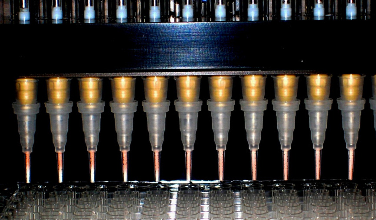
The probe syringe format overcomes the main disadvantages of the spin format, including limited control of the unidirectional flow rate and a more complex automation process in cases of multistep workflows. For this format, a robotic liquid-handling head is fitted with special syringes, each coupled directly to liquid-handling probes, as shown in Figure 3. The tip of the probe fits into the inlet seal of the cartridge, forming a direct liquid connection between the inside of the syringe and the packed bed. The seal taper is small enough so that it can hold a liquid pressure of at least 15 bar with just friction. A broad range of flow rates through the cartridge can be achieved, from well under <1 to >5000 μL/min. The syringe plunger also has a tapered shape that tightly fits the inside of the probe, providing an ultra-low dead volume in the syringe, which both effectively ejects air bubbles and enables efficient washout between samples and reagents. The probe is also designed so that a standard pipette tip can be attached, enabling the same liquid-handling system to perform both cartridge operation and conventional liquid handling. The probe syringes are mounted in a modified version of a standard 96-channel pipetting head, so that a full 96-well microplate of samples can be processed in parallel. A commercial system (AssayMAP Bravo 96AM; Agilent Technologies, Santa Clara, CA) has been introduced which implements this technology.
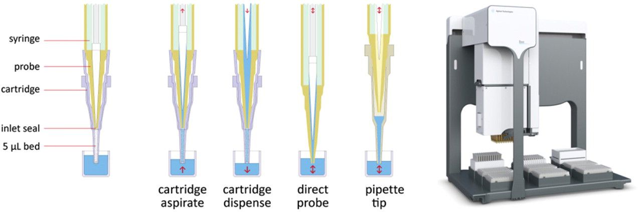
Probe syringe format, showing the elements of the ultra-low dead volume probe syringe and its connection to the cartridge. This system enables highly precise flow control through the cartridge in either direction, as well as both direct and pipette tip-based liquid handling with the same head. The commercial Agilent Technologies AssayMAP Bravo 96AM with a 96-channel array of probe syringes is shown.
The probe syringe is able to precisely aspirate and dispense fluids either directly or when attached to the cartridge. With upward aspiration, a series of different liquids can be pumped through the cartridge bed in sequence, without disconnecting the cartridge and washing out the syringe in between. Downward dispensing is used for steps where the eluate is to be recovered into a microplate well and can also be used to apply high pressures to the bed. Samples can also be aspirated and dispensed in a rapid cycle to distribute binding evenly throughout the bed, which can be useful in immobilization or coating steps.
Affinity Purification and Quantitation
Perhaps the most basic application of the microchromatography platform is to use an affinity ligand immobilized on a resin in the cartridge to selectively bind the target protein. After washing away the unbound impurities, the purified target protein is eluted and recovered in a microplate well. The amount of target protein recovered from the packed bed cartridge can be precisely and accurately quantitated in the microplate well, using methods such as UV absorbance at 280 nm or protein assay reagents such as Coomassie Blue on standard plate readers. Because binding to and elution from the cartridge is itself quantitative, this simple method can be used to accurately quantitate the target protein in the original sample. Figure 4 shows data in the spin and probe syringe formats for quantitation of monoclonal antibodies in complex sample matrices such as cell culture supernatant using protein A as an antibody-specific affinity ligand. Both absolute quantitation and analytical precision of this method are very similar to protein A HPLC (the standard method used in the biopharmaceutical industry), but because many samples can be run in parallel, the throughput is much higher.
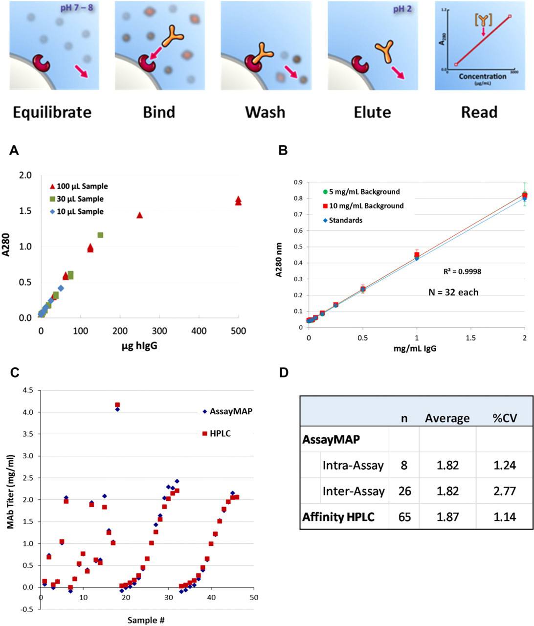
Purification of IgG antibodies using protein A affinity cartridges. (A) IgG standards run in the spin format at various concentrations in different sample volumes. When plotted as A280 nm versus IgG mass, all the curves overlay, indicating quantitative binding up to approximately 100 μg per cartridge. (B) Linear range runs on a probe syringe system. 5 IgG spiked into buffer and high concentrations of background protein (fish gelatin) gave identical results, indicating good purification. (C) Comparison of cell culture supernatant samples of a monoclonal antibody produced in a Chinese hamster ovary-cell bioreactor analyzed by protein A HPLC and AssayMAP protein A cartridges run on a Tecan EVO automation system (Tecan Group, Ltd., Mannedorf, Switzerland). 6 Concentrations of antibody in the samples were determined by taking the absorbance of the proteins eluted from the HPLC or the AssayMAP cartridge and comparing the absorbance to a standard curve. (D) Quantitative comparisons of AssayMAP cartridges and protein A HPLC show very similar results and precision.
In addition to quantitation, the recovered target protein is also available in purified form for additional analytical methods. The amount recovered for an individual sample typically can range from 1 to 100 μg for cartridges with a 5-μL packed bed, which is sufficient for many analytical applications.
The affinity ligands used in the cartridges can include protein ligands, antibodies and antigens, cell surface receptors, or synthetic chemical ligands (such as are used in immobilized metal affinity chromatography to target his-tagged proteins). The ligands can be preimmobilized on the resin before it is packed or, more preferably, immobilized in situ, using either hydrophobic adsorption or methods such as biotin/streptavidin binding. Figure 5 shows an example of a human -specific camelid VHH-binding domain protein 7 immobilized in cartridges used to quantitatively purify human IgG from rat serum (as would be required in a preclinical pharmacokinetic study). Protein A is not suitable for this application because it binds the abundant rat IgG in the sample along with the human IgG.
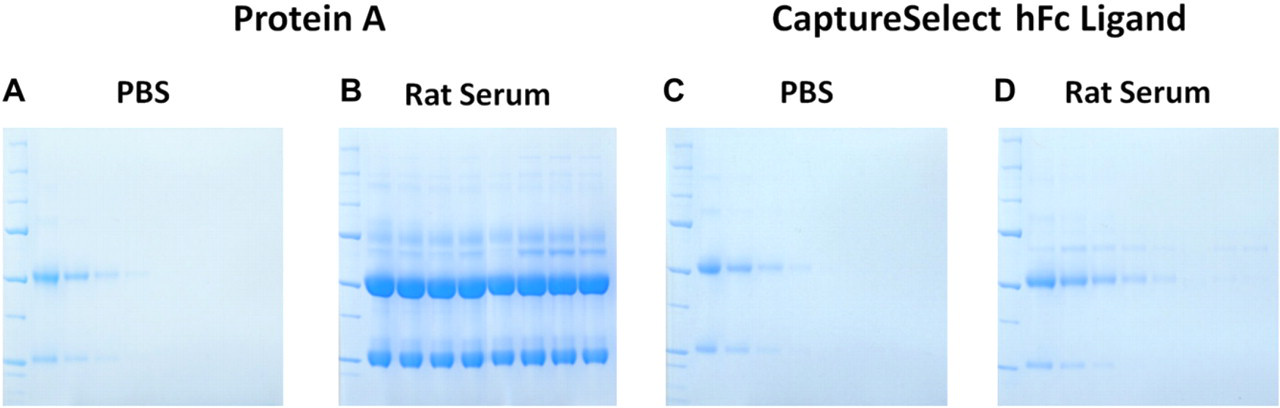
Human IgG was spiked either into phosphate-buffered saline buffer (A and C) or normal rat serum (B and D) at amounts of 20, 10, 5, 2.5, 1.25, 0.63, 0.31, and 0 μg and loaded on to cartridges with either protein A resin (A and B) or CaptureSelect hFc (BAC, BV, Naarden, The Netherlands) resin (C and D). CaptureSelect hFC resin contains a camelid antibody VHH-binding domain protein selective for human IgG over most animal IgGs. The cartridges were loaded, washed, and eluted using standard protocols in the spin format (see cartoon in Fig. 4), and the eluted fractions run on sodium dodecyl sulfate polyacrylamide gel electrophoresis gels stained with Coomassie Blue. The CaptureSelect hFc cartridge could selectively and quantitatively bind human IgG from the rat serum, whereas the protein A cartridge showed cross-reactivity with the rat IgG.
Immunoassay
AssayMAP cartridges can also be used to perform a number of different types of immunoassays, including enzyme-linked immunosorbent assays (ELISAs). For these applications, the cartridge is packed with large-particle nonporous beads with a hydrophobic surface. The total surface area in the cartridge is similar to a standard microplate well, used as the solid phase in many immunoassay procedures, and similar antibody immobilization methods (such as passive hydrophobic immobilization or biotin–streptavidin) may be used. However, because of the packed bed format, the diffusion path for the molecules in the sample to reach the binding ligands on the solid-phase surface is much shorter—just the micron-scale space between the beads versus the millimeter-scale size of the microplate well. As a result, binding reactions go to completion much faster, eliminating the need for long incubation steps. A standard sandwich ELISA can be completed (from coating to readout) in 30 min versus the 4–24 hours required for standard microplate methods, with no change in the reagents, buffers, or standards used.
Immunoassays on the system start with the coating step, which typically takes just a few minutes; so precoating and subsequent storage of cartridges is not required. After blocking, the sample is loaded at a low-enough flow rate to enable complete binding (typically around 5 μL/min). For maximum sensitivity, it is often advantageous to preincubate the sample with the labeled conjugate antibody; but in other cases, the conjugate antibody is loaded on the cartridge after the sample. After washing (which is very rapid and efficient because of the packed bed format), the syringe is filled with enzyme substrate solution, which is dispensed through the cartridge, and the resulting product is collected in a microplate for readout on a standard plate reader. The precise control of flow rate is critical for this step because the amount of product formed (and thus the signal) is inversely proportional to the residence time of the substrate solution in the packed bed.
Figure 6 shows typical results with an ELISA for protein A in solution (a common assay in the bioprocess industry, where protein A is a possible product contaminant). Reagents from a commercial microplate assay kit were run in both the standard microplate format and on the AssayMAP cartridges. Overall results were nearly identical, with coefficients of variation for the assay well under 10%. Note that at the low end of the range, the total protein A target quantitated was 10 pg.
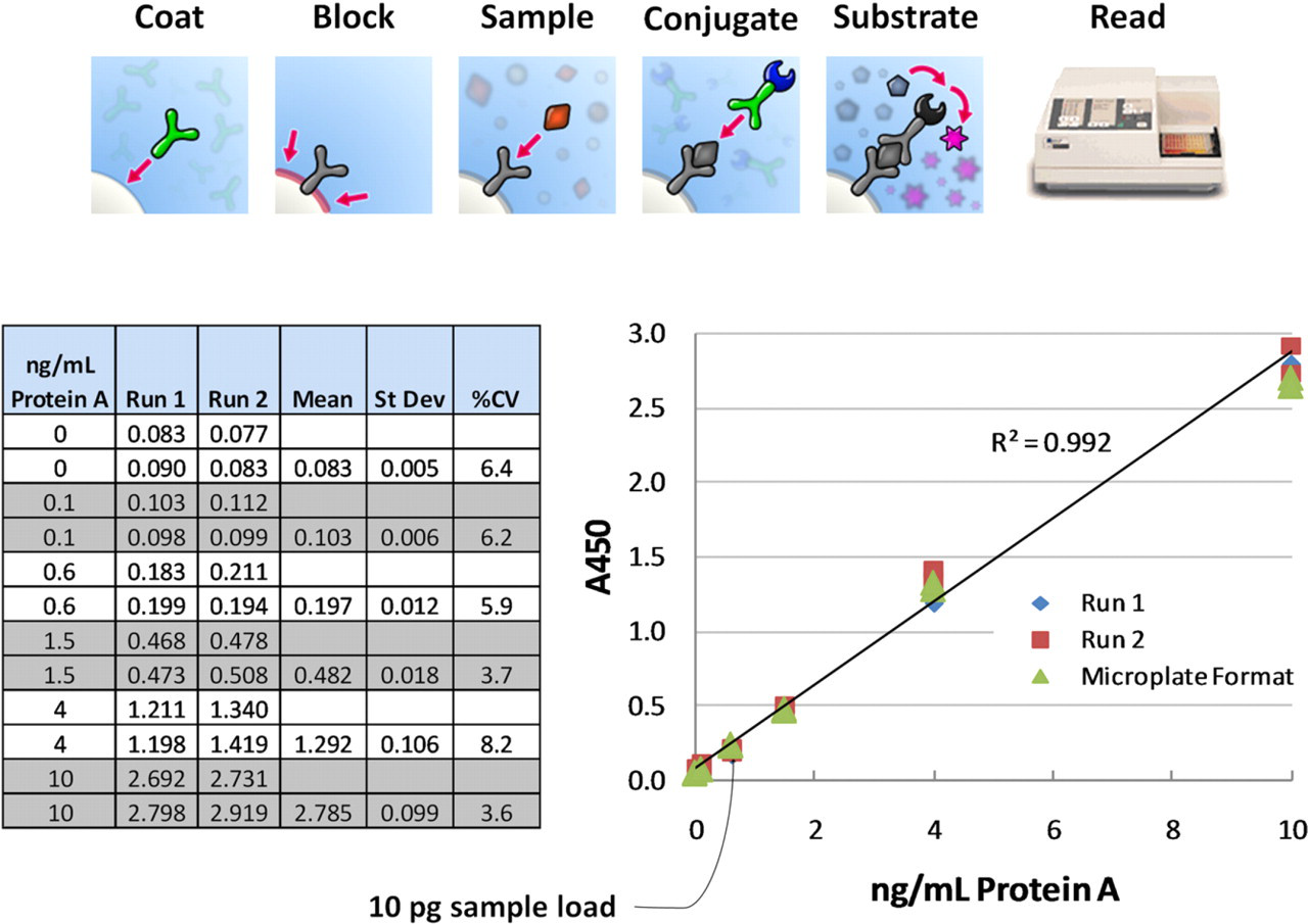
Reagents from a commercial ELISA kit for protein A in solution (Cygnus Technologies, Southport, NC) were run on AssayMAP ELISA cartridges containing nonporous beads with a hydrophobic binding surface, similar to a polystyrene microplate. The plot compares the results from the assay kit package insert (run in the standard microplate format) with the results for the same standards in the cartridge format. The cartridge process took 30 min per run versus 4 h for the microplate method.
Glycan Analysis
Glycan analysis is an example of sample preparation for more complex structural analysis of proteins. The profile of glycoforms found in a biotherapeutic protein can be quite complex and can significantly affect the safety and efficacy of the drug; yet control of the cell culture conditions to produce a particular glycan profile is poorly understood. 8 In addition, the demand for glycan analysis is increasing dramatically, but sample preparation for glycan profile analysis is typically a laborious manual method. 9 Only a few 10s of samples are usually processed at a time, and the entire procedure can take up to 3 days.
High-throughput microchromatography can be used to dramatically speed up and automate glycan sample preparation. The target glycoprotein is first purified from complex samples, using the affinity purification methods described above. The target glycoprotein is then immobilized on resin in a cartridge and exposed to an enzyme such as PNGase F, which selectively cleaves off the N-linked glycan groups. The released glycans are eluted from the cartridge and collected in a well in which they are reacted with fluorescent labels for sensitive analysis. In some methods, the labeled glycans are then bound to a cleanup cartridge, excess label washed away, and the cleaned up labeled glycans are eluted for analysis by HPLC, LC-MS, or capillary electrophoresis. These workflows can be performed using either the spin or the probe syringe formats.
Figure 7 A and B compare the resulting glycan analysis profiles (by hydrophilic interaction (HILIC) HPLC with fluorescence detection) for two typical glycoproteins prepared by the cartridge-based method (GlykoPrep system; ProZyme, Inc.) described above and conventional methods. The retention times for the GlykoPrep samples are somewhat longer because the labeling chemistry used (which is extremely rapid and eliminates the need for sample drying steps) is slightly more hydrophilic than the conventional chemistry. However, the overall profiles are virtually identical. Figure 7C and D illustrate the excellent reproducibility of the cartridge-based method.
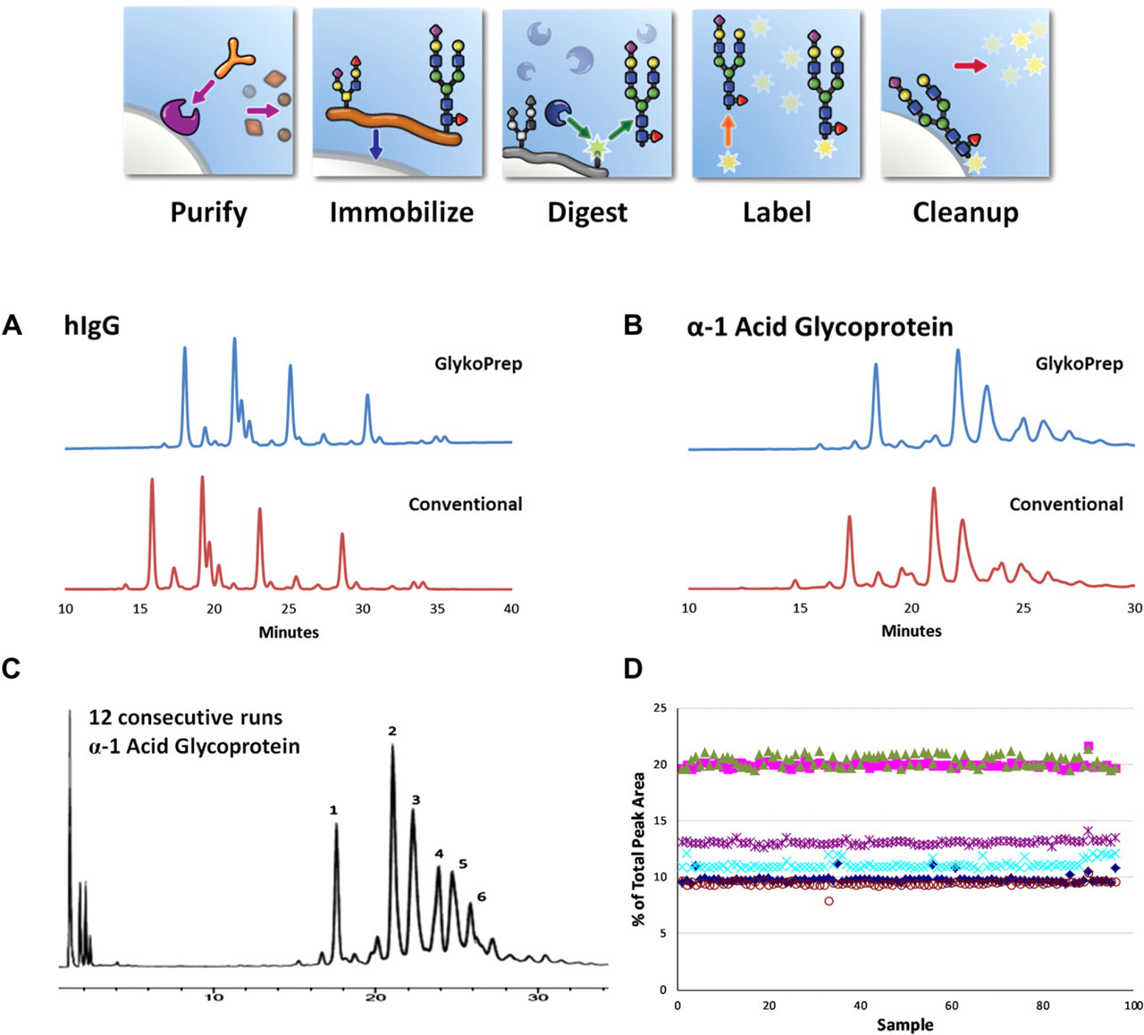
(A) and (B) show hydrophilic interaction (HILIC) HPLC chromatograms with fluorescence detection for N-linked glycan samples from human IgG and human α-1 acid glycoprotein, respectively. 10 Samples were prepared using either the GlykoPrep cartridge-based method (ProZyme, Hayward, CA) or standard methods with 2-aminobenzamide labeling. (C) Twelve overlaid chromatograms from an entire 96-well plate of identical human α-1 acid glycoprotein samples and (D) shows the percentage total peak area of the six major peaks for all 96 samples, illustrating the high reproducibility of the method.
Chromatographic Method Development
Because AssayMAP cartridges function as true chromatography columns, it is possible to use the platform as a tool for chromatographic method development, with the benefits of being able to run many operating conditions in parallel using relatively small amounts of sample. One potential application is to screen pH and ionic strength as independent variables for the washing and elution conditions of ion exchange chromatography separations. Figure 8 shows a cation exchange separation of cytochrome C from ribonuclease A at a particular pH, following a wash with different salt concentrations from 0 to 100 mM. The cytochrome C contains a red-colored heme group, so it is visible in the packed bed. At low ionic strength (far left), the protein is tightly bound to the top of the bed after the wash, whereas at the high ion strength (far right), almost all of the protein is washed off the bed. There is a continuous gradation at intermediate ionic strengths. This suggests that despite its very small bed volume, the cartridge system is capable of good chromatographic performance.
A row of 12 cartridges packed with cation exchange resin and loaded with a mixture of cytochrome C and ribonuclease A. After sample loading, the cartridges were washed with 25 μL (five bed volumes) of buffer at pH 6.5, with a series of linearly increasing concentrations of NaCl from 0 (far left) to 100 mM (far right). The cytochrome C has a red-colored heme group so that its binding behavior can be clearly seen on the cartridge beds.
A different application is illustrated in Figure 9. In these experiments, the probe syringe was filled with a feed stream containing a binding protein (in this case IgG), cartridges were loaded, and the feed stream pumped through at a particular flow rate. The output of the cartridge was collected in very small volume fractions in adjacent wells of a microplate. The breakthrough curve of binding protein is determined by adding Bradford protein analysis reagent and reading the protein concentration of the collected fractions with a plate reader. In this case, several different resins were run in parallel to determine their dynamic breakthrough capacity as a function of flow rate (or residence time, equal to the cartridge bed volume divided by the flow rate). Two different resin types are shown at three different flow rates. The results were very similar to those obtained for the same resins and feed stream in a conventional system with a 1-mL column, despite the 200-fold reduction in volume and flow rate.
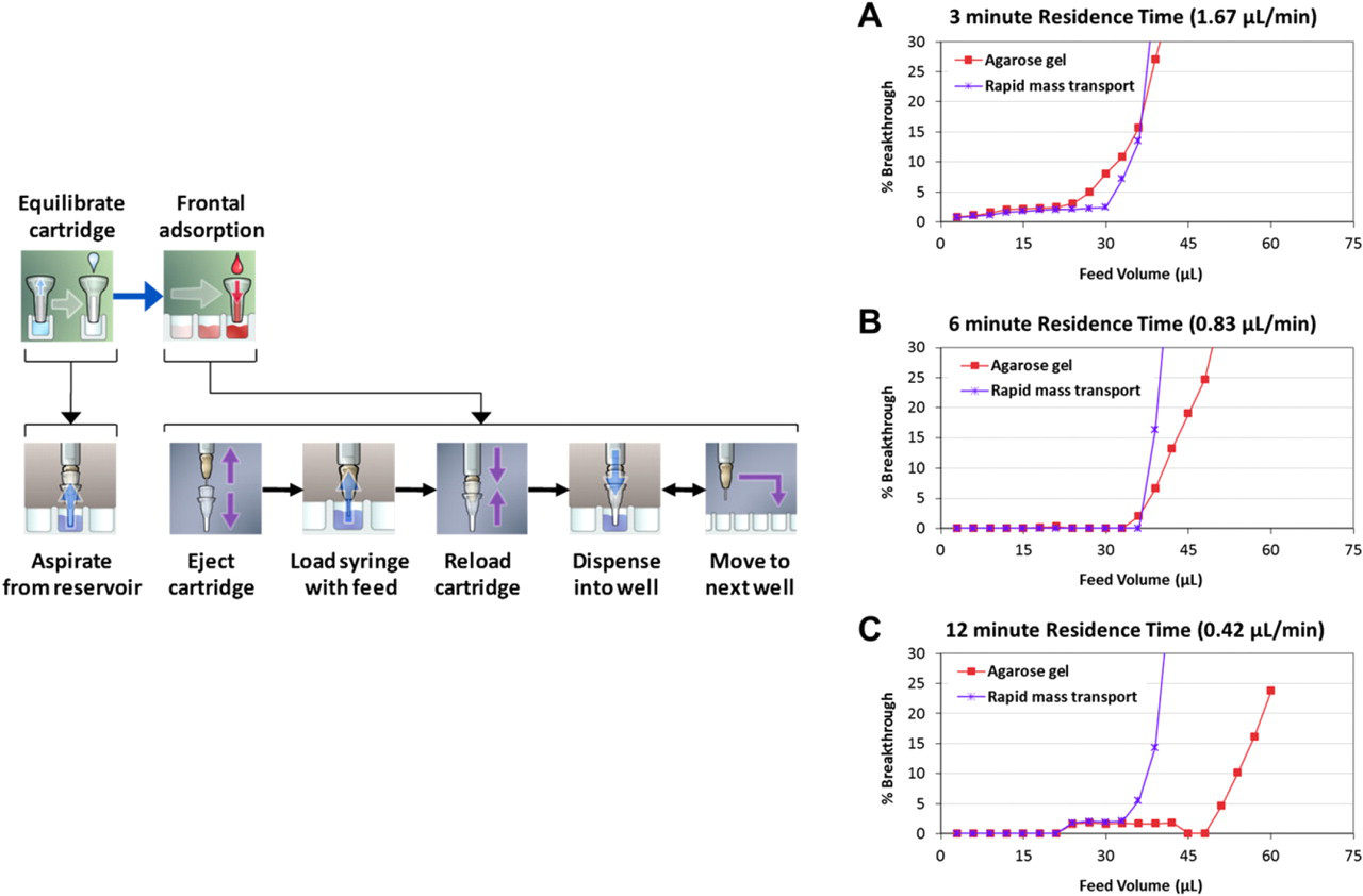
Frontal adsorption analysis of IgG binding in cartridges packed with two different protein A resins at three different flow rates (A: 1.67, B: 0.83, and C: 0.42 μL/min). 11 Small (3 μL) fractions were collected in adjacent wells of a microplate and Bradford Coomassie Blue reagent was added to measure the breakthrough protein concentration relative to the feed concentration by absorbance.
Summary
The biotherapeutic industry is driving an ever-increasing demand for protein analysis. Chromatography is a well-proven and effective general method to purify, quantitate, and enzymatically react protein samples, but meeting the technical challenges of scale, throughput, quantitative binding and elution, and so forth has been difficult. The AssayMAP high-throughput microchromatography system can provide quantitative chromatographic separations in either a spin format (requiring no special equipment) or a probe syringe format (using a special liquid handler, but providing precise bidirectional flow rate control), both compatible with microplate-based automation. Applications for general protein purification and quantitation, chromatographic method development, and immunoassay have been demonstrated, along with complex workflows involving multistage purification and enzymatic digestion for glycan profile analysis.
Footnotes
Acknowledgments
Financial support for this work was provided by BioSystem Development, LLC. The authors would like to thank Michael Bovee for a detailed review of the manuscript.
Competing Interests Statement: All authors of this article are employees of the supporting institution, BioSystem Development. Scott Fulton, Zachary Van Den Heuvel, Ronald Smith, and Robert Sakowski are also stockholders of the company.