
Abstract
A fluorescence resonance energy transfer (FRET)- based aptamer biosensor (aptasensor) for sensitive determination of adenosine triphosphate (ATP) was developed. The energy donor and acceptor were 5′-carboxyfluorescein (5′-FAM) and gold nanoparticles (AuNPs), respectively. AuNPs were linked to 3′-SH-DNA by Au-S bond. This aptasensor was obtained by the hybridization between the complementary DNA strands of the 5′-FAM and AuNPs. The detecting results indicated that the FRET increase efficiency (Fr) of detecting system at 519 nm was linear when the concentration of ATP target was about 0.1–10 mmol/L. Moreover, the aptasensors showed a good stability in different buffer conditions and a low- detection limit (15.2 nmol/L) for ATP.
1. Introduction
Adenosine triphosphate (ATP) is a fundamental biological energy carrier in cells, and many cellular processes are modulated by the extracellular ATP. Moreover, the viability and injury of cells can also be indicated by ATP [1–2]. Therefore, the extracellular or intercellular detection of ATP gains great interests in biosensor field [3], for instance, fluorescent, electrochemical, and colorimetric biosensors [4-6]. Compared with other methods, fluorescence detection has high sensitivity, low cost, good specificity, and other advantages to develop rapidly.
Nowadays, aptamers, single-stranded nucleic acids, get a great improvement. They can be artificially selected from DNA libraries, chemically synthesized, and have selective and special binding to many targets, even small molecules [7]. Therefore, the aptamer biosensor (aptasensor) can be easily obtained and applied in various analysis fields [8, 9], such as bacteria [10], small molecules [11], and drugs [12]. Although the aptamer and target have strong affinity, the sensitivities of reported ATP aptasensors were not low enough [13-15]. Zhao et al. [13] reported an aptamer fluorescence anisotropy sensor for ATP detection when tetramethylrhodamine (TMR) was as the fluorescent label, and the detection limit was of 1 mmol/L. Zhang et al. [14] prepared an Ag/SiO2 core/shell nanoflare aptasensor based on metal-enhanced fluorescence for ATP detect when cyanine as the fluorescent label, and the detection limit was about 8 × 10⁻⁶ mol/L. Wei et al. [15] prepared a fluorometric ATP aptasensor with exonuclease I as digesting nuclease and no label. The detection limit was about 0.14 × 10⁻⁶ mol/L.
The fluorescent biosensors designed by fluorescence resonance energy transfer (FRET) process should exhibit a low- detection limit about 10−12–10−9 mol/L because inorganic nanoparticles, quantum dots (QDs), and AuNPs are utilized in this field [16, 17]. Usually, AuNPs are excellent energy acceptors by virtue of their good bioinert and quenching ability [18]. According to FRET mechanism, there should be an overlap between the ultraviolet-visible (UV-vis) spectra of energy acceptor and fluorescent emission spectra of energy donors. Wang et al. [19] reported a FRET-based aptasensor of Ochratoxin A (OTA) when fluorescein amidite (FAM) and gold nanoparticles (AuNPs) were as energy donor and acceptor pair, respectively, and the detection limit was about 2 × 10⁻¹² g/mL. Sun et al. [20] presented a FRET-based aptasensor of recombinant human erythropoietin-α (rHuEPO-α) when AuNPs were as the energy donors, and the detection limit was about 0.92 × 10⁻⁹ mol/L.
In this chapter, a FRET-based aptasensor for ATP detection with high sensitivity and selectivity is presented. The mechanism was shown in Figure 1. The 5′-carboxyfluorescein (5′-FAM) and AuNPs were as the energy donors and acceptors and conjugated with two complementary single-stranded DNAs (ssDNAs), respectively. When the donor and acceptor were close enough (within 1–10 nm) after the hybridization of ssDNA, the FRET process should occur from donor to acceptor and resulting in the quenching of the fluorescent intensity of aptasensor. If the ATP as target existed in detection system, the double-stranded DNA (dsDNA) structure of aptasensors would be opened because of the stronger hybridization between the ATP and the aptamer ssDNA, as well as the fluorescent intensity of detection system could be increased.

The principle of detection of ATP aptamer probe
2. Experiment
2.1 Reagents and instruments
HAuCl4·4H2O were from Kermel Chemical Reagent Company (Tianjin, China). Tris(hydroxymethyl)-aminomethane was obtained from Alfa Aesar. ATP, uridine triphosphate (UTP), guanosine triphosphate (GTP), and cytosine triphosphate (CTP) were purchased from Sigma-Aldrich. All other reagents were of analytical grade. The oligonucleotides, ATP aptamer (DNA1), and its complementary strand (DNA2) were supplied by the SBS Genetech Company (Beijing, China). The base sequence of the DNA1 was 3′-SH-(CH2)6-TGG AAG GAG GCG TTA TGA GGG GGT CCA-5′, and the sequence of DNA2 was 3′- AA CGC CTC CTT CCA-FAM-5′.
2.2 Preparation of AuNPs
The Fren's method was utilized to obtain AuNPs of 16 nm in diameters when the citrate was as the reduction [21]. HAuCl4 (1 mL, 1%) was added into 100 mL of doubledeionized water and fluxed till boiling. Then, trisodium citrate solution (2.5 mL, 1 %) was quickly added into this mixture with strong stirring for 15 min. After the color of reaction system became wine red, the reaction should be stopped and cooled to room temperature. The AuNPs were separated and purified by centrifugation (10,000 r/min about 40 min), resuspended in aqueous solution (100 mL), and stored at 4°C. The UV-vis absorption spectra and transmission electron microscopy (TEM) of AuNPs were measured by Helios-γ thermo ultraviolet and visible spectrophotometers (USA) and JOEL JEM-2100F FasTEM (Japan), respectively.
2.3 Self-assembly of DNA1 on AuNPs surface (Au-DNA1)
DNA1 and AuNPs were incubated at room temperature for about 16 h when 3′-SH-DNA (DNA1, 1OD) was mixed with AuNP suspension (15 mL). Then, the solution was transferred into a 10 mmol/L of phosphate-buffered saline (PBS) solution with 0.1 mol/L of NaCl (pH = 7.0) under shake at 37 °C. After 40 h, the reaction could be ended, and DNA1 conjugated AuNPs (Au-DNA1) obtained. The resultant Au-DNA1 conjugates were purified by ultracentrifugation of 10,000 r/min with 0.1 mol/L of NaCl/PBS and resuspended in PBS.
2.4 Preparation of FRET-based aptasensors
The aptasensor probe was obtained after the hybridization of Au-DNA1, and FAM-DNA2 was incubated in a Tris-HCl (20 mmol/L) with 150 mmol/L of NaCl buffer solution at 37°C for about 12 h. The detection process was that an amount of ATP was introduced into the aptasensor detection solution under shake. After 12 h, the fluorescent spectra of the detecting solution were measured by F-7000 fluorescence spectrophotometer (HITACHI, Japan). The FRET increase efficiency (Fr) was given as follows:

where F0 was the fluorescent intensity of the detecting system, as well as F was the fluorescent intensity of detection system with ATP target.
3. Results and Discussion
3.1 Self-assembly of DNA1 with AuNPs (Au-DNA1)
The TEM result of AuNPs was shown in Figure 2, which indicated that the AuNPs had a uniform size; the diameter was about 16 nm, as well as the particle shape was spherical; and the dispersion was even [22].
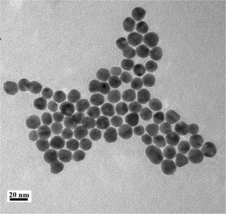
TEM image of Au nanoparticles
The AuNPs could be self-assembled by DNA1 due to the strong affinity of Au-S bond. Au-DNA1 precipitates were investigated by UV-vis spectra when molar ratios of AuNPs and DNA1 changed after 10,000 r/min centrifugation (Figure 3). DNA cannot be precipitated at this centrifugation speed [23]. Therefore, the free DNA1 should be in the supernatant and be removed after centrifugation. The results of Figure 3 indicated that the Au-DNA1 conjugates had the characteristic absorption peaks of AuNPs and DNA at 520 and 260 nm, respectively. The peak of 260 nm had a gradual increase when the amount of DNA1 was increased, which indicated that the AuNPs carried more DNA1 on their surfaces. However, the increase in tendency of absorption peak at 520 nm of AuNPs weakened gradually. The UV-vis spectrum of AuNP aqueous dispersion is from the surface plasmon resonance of particles. Therefore, particle size and Au molar concentration can be measured directly from UV-vis spectra [24]. Moreover, the increase in UV-vis intensity can also indicate the dispersion improvement of lyophobic AuNP systems when the amount of AuNPs is fixed [25], which resulted that the UV-vis spectra of Au:DNA1 = 1:100 and 1:150 had only a very slight change at 520 nm of AuNPs but an obvious change at 260 nm of DNA1.
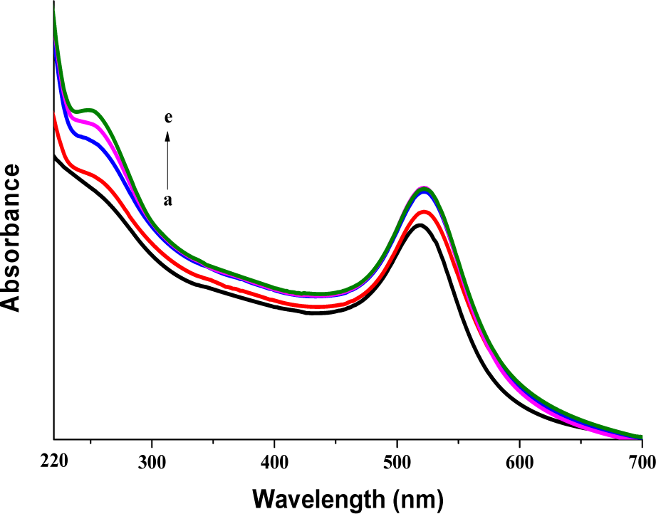
Influence of molar rations on Au-DNA1 precipitates when the concentration of AuNPs was about 2.3 nmol/L: (a) Au; (b) Au:DNA = 1:10; (c) Au:DNA = 1:50; (d) Au:DNA = 1:100; (e) Au:DNA = 1:150
3.2 Construction of aptasensor system
According to the principle of FRET, the acceptor and donor should have a reasonable distance (about 1 −10 nm) and a certain overlap of the UV-vis absorption spectrum of the acceptors and the fluorescent emission spectrum of the donors [26]. In this case, the AuNPs could quench the fluorescence of 5′-FAM because the distance was close enough by the complementary hybridization of ssDNA. The result of the two spectra was shown in Figure 4, which demonstrated that there was a large overlap at 519 nm. Therefore, the Au-DNA1 and FAM-DNA2 were suitable donor and acceptor for aptasensor based on FRET theory. The formation of fluorescent aptasensors (FAM-dsDNA-Au) was by the hybridization of 5′-FAM-DNA2 and Au-DNA1. The acceptor, AuNPs, quenched the fluorescence of donors, 5′-FAM, and resulted in the energy transfer [27]. The fluorescent changes of 5′-FAM-DNA1, aptasensors, and the aptasensors with ATP target were shown in Figure 5. The fluorescence of 5′-FAM was quenched strongly after the aptasensor formation, which indicated that the distance between FAM and AuNPs could be within 1–10 nm after hybridization, and the FRET (quenching) efficiency was about 76%. When ATP target existed, the hybridized structure of aptasensor had to be opened. This would result in the departure of FAM-DNA2 from the aptasensor structure and the Fr of detection system (about 53.5%). Therefore, this aptasensor system could detect the existence of ATP.

Spectra overlap of (a) fluorescent emission of 5′-FAM and (b) UV absorption of AuNPs

Construction of fluorescence resonance energy transfer system: (a) FAM-DNA; (b) Au-DNA1; (c) probe; and (d) probe + ATP
3.3 Detection ability of the fluorescent aptasensors
The sensitivity of this aptasensor system was investigated under different ATP concentrations (Figure 6). The fluorescent intensity showed a gradual increase with the addition of ATP concentration. When the ATP concentration range was 0.1–10 mmol/L, the corresponding linear regression equation was Fr = 0.263c at 519 nm with R2 of 0.996, where Fr and c were the Fr and logarithm of ATP concentration, respectively. The detection limit for ATP was about 15.2 nmol/L.

Detection of ATP with different concentrations and the corresponding working curve: (a)–(g) no ATP, 0.1 μmol/L, 1 μmol/L, 10 μmol/L, 0.1 mmol/L, 1 mmol/L, and 10 mmol/L when the concentration of AuNPs was about 2.3 nmol/L
The selectivity and specificity of the ATP aptasensor were investigated when the analogs, UTP, CTP, and GTP, were introduced (Figure 7) due to the fact that they had similar molecule structures with ATP and could interfere with the detection in practical applications. The results indicated that when the concentration of three analogs was 10- folds to ATP concentration (1 mmol/L against 100 μmol/L), only ATP had an obvious recovery of fluorescent intensity. The presence of ATP analogs had nearly no fluorescent influence, which indicated that ATP analogs could not open the hybridized dsDNA structure of ATP aptasensors. Therefore, this ATP aptasensor had a good specificity.
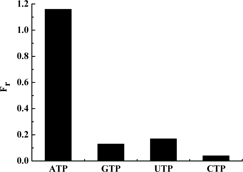
Selectivity of the aptasensor when the ATP concentration was about 100 μmol/L, and the GTP, UTP, and CTP concentrations were about 1 mmol/L
3.4 Stability of the fluorescent aptasensors for ATP
Both the fluorescence of donors and the scaffold structure of dsDNA could have some changes by the buffer conditions [13, 28]. Therefore, the stability of this aptasensor system was investigated in different buffer conditions and pH values (Figure 8). The results indicated that the Fr of the detection system reached the maximum when pH value was 8, and the ATP concentration was 100 μmol/L (Figure 8a). Moreover, the fluorescent background intensity of FAM would be increased greatly when pH value was more than 8 [28]. Therefore, the reasonable pH for detection of FAM- based biosensors was about 7.5. The effect of NaCl on Fr in the binding buffer solution indicated that the Fr did not show obvious changes under different NaCl concentration (Figure 8b), which was also confirmed by Zhao et al. [13].

The fluorescence response of ATP aptasensor in different buffer conditions with 20 mmol/L Tris-HCl: (a) pH and (b) NaCl when the ATP concentration was 100 μmol/L
4. Conclusions
An aptamer scaffold and FRET- based fluorescent aptasensor for ATP detection were presented in this chapter when 5′-FAM and AuNPs were as donors and acceptors, respectively. The donors and acceptors were conjugated with complementary ssDNA. The fluorescence of donors could be quenched by acceptors because of the hybridization of ssDNA each other in different buffer solutions. When ATP as target existed, the Fr of this detection system was gradually increased. While the targets were other small molecules, UTP, CTP, and GTP, the Fr had no significant changes. The relationship of Fr and ATP concentration was linear within the ATP concentration from 0.1 to 100 μmol/L, and the detection limit was about 15.2 nmol/L.
Footnotes
5. Acknowledgements
The work was supported from the National Natural Science Foundation of China (Grant Nos. 21172171 and 21206124) and Natural Science Foundation of Tianjin (Grant No. 15JCYBJC20500).