
Abstract
Highly monodispersed CdS nanoparticles using polyvinyl pyrrolidone (PVP) as the capping agent were synthesised by chemical coprecipitation method. The surface-modified cadmium sulfide nanoparticles were found to be remarkably stable. In the presence of PVP, cubic phase with small grain size of CdS were observed in XRD. The peaks were identified to originate from (111), (220) and (311) planes of CdS, respectively. The crystallite size of the synthesised CdS nanoparticles was about 3 nm calculated from the (111) plane of XRD pattern and it was also confirmed through HRTEM. Morphology and elemental mapping of the synthesised nanoparticles were studied by SEM and EDX analyses. Increase in the band gap with decrease in the particle size was observed from the reflectance mode UV spectrum, which confirms the quantum confinement effect. From the photoluminescence studies, enhanced near-band-edge blue light emission and significantly reduced defect-related green emission were observed. Longitudinal optical (LO) phonon modes, corresponds to pure CdS were observed in Raman spectrum.
1. Introduction
Semiconductor nanocrystals have attracted impressive attention because of their novel optical and electronic properties. Direct and wide bandgap semiconductor nanomaterials are potential candidates for applications in nonlinear optics and optoelectronics [1]. Varying the size of the particle changes the degree of confinement of the electrons and affects the electronic structure of the solid, in particular band edges, which are tunable with particle size.
Efficient luminescent quantum dots form an important and interesting class of luminescent materials. Their broad absorption spectrum and narrow emission band would be tunable by changing their size. They have demonstrated excellent optical properties and higher photochemical stability than most organic emitters [2]. Most of the physical and chemical properties exhibited by these nanoparticles are due to their crystallite size. Further growth in the size is due to agglomeration of these crystallites to form bulk particles. If this growth of the particle is not controlled, then due to Ostwald ripening and Vander Waals interactions between particles, they get agglomerated [3–4]. This agglomeration can be avoided by stabilizing them electrostatically at appropriate stages to achieve size selective synthesis during precipitation reaction.
Due to its large surface to volume ratio, the atoms situated near the surface regions play a major role in its electronic, optical and thermodynamic properties. At the surface, since the co-ordination number around the atoms is less than that inside the bulk, there can be larger number of defect states which acts as non-radiative pathways for the excited electrons and become detrimental to the luminescent properties of nanocrystalline phosphors [5]. Therefore, it is necessary to modify the nanoparticle surface by using suitable capping agents so that the surface caps will passivate the defect states and dangling bond density. Covering the nanocrystal core surface with an inorganic shell or organic ligand molecules can bring about necessary passivation of vacancies, stabilization of the colloidal suspension and also maintain the quantum confinement effects by way of particle isolation. Several methods have been used to improve the stability of nanoparticles, such as changing of annealing temperature, doping of semiconductor and surfaces capped by various organic or inorganic layer etc. [6–8]. Among these methods, polymer capping in a chemical method has been developed to synthesize nanoparticles with highly surface stability, and also has significant influence on the morphology and optical properties of nanoparticles [9]. Various wet chemical methods have been developed for the synthesis of sulphide nanoparticles [10–12].
As a direct band gap semiconductor with Eg of 2.42 eV at room temperature, CdS nanostructural materials have been prepared using various physical and chemical solutions with a view of their commercial or potential applications in light-emitting diodes, solar cell and optoelectronic devices [13–14]. The size of the quantum dots obtained by this method can be controlled by capping the nanoparticles using (polyvinyl pyrrolidone) PVP as a stabilizing agent. In this work, we have studied the structural and optical properties of PVP capped CdS quantum dots synthesized by the coprecipitation method.
2. Experimental techniques
2.1 Synthesis of PVP capped CdS nanoparticles
Starting materials for the synthesis of CdS nanoparticles were cadmium acetate dihydrate, sodium sulphide and poly-n-vinyl-2-pyrrolidone (PVP). All the reactants were 99.9% pure and used without purification. PVP was used as the dispersant that adsorbs the single colloidal particle to form a molecular folium to prevent the particle from coalescing.
Aqueous solution (30 ml) of 0.35 M cadmium acetate and 0.35 M sodium sulphide was prepared separately with DI water (resistivity 10−18 mΩ). Sodium sulphide solution was added dropwise to the aqueous cadmium acetate solution, under stirring condition. After 5 minutes of stirring, 0.5 g of PVP was added under vigorous stirring for 2 h at room temperature. pH of the reaction mixture was adjusted to ∼11 by dropwise addition of aqueous ammonia solution. The mixed solution was then refluxed for 1 h at constant temperature of 70°C. After cooling to room temperature the obtained precipitate was ultrasonically treated for 20 minutes. The color of the solution appeared to be bright yellow indicates the formation of cadmium sulphide. The sample was then washed and filtered with DI water and ethanol several times to remove the excess organic residues. The collected sample was dried and stored in the desiccator for further characterization. For comparison pure CdS also synthesised without PVP capping in the above same procedure.
2.2 Characterization of the CdS nanoparticles
X-ray diffraction pattern were recorded using PANalytical X-ray diffractometer with CuKα radiation (λ = 1.5406 Å) in the range of 20°–60° (2θ) at a scanning rate of 0.05°/min. Hitachi S-4800 HR-FESEM and EDX with an acceleration voltage between 10 and 15 kV were used to analyse the morphology, elemental analysis and mapping of the synthesised nanoparticles. High resolution transmission electron microscope and SAED pattern were performed at 200 keV using JEOL JEM 3010 with LaB6 filament. UV-reflectance studies were carried out using CARY 5E UV-VIS Reflectance mode spectrophotometer. FT-IR spectra were recorded by Perkin Elmer Spectrometer. Photoluminescence spectra were recorded using Shimadzu-5301 spectrophotometer. Raman measurement were analysed with Horiba Jobin-Yvon T64000 micro-Raman spectroscopy with Ar laser source at a laser power of ∼50 mW with the excitation wavelength of 514.5 nm.
3. Results and Discussion
3.1 Structural analysis
Powder X-ray diffraction of synthesised nanoparticles (Fig.1) shows a perfect match with the cubic zinc blende phase of CdS (JCPDS 10-454). The diffraction peaks of the nanoparticles are considerably broadened due to the small size of the crystallites. The peaks can be indexed as (111), (220) and (311) which are characteristic peaks of crystal planes for CdS cubic phase. It has been observed that the surface capping with PVP molecule does not have any effect in the crystal structure of CdS nanoparticles. The crystallite size of the synthesised PVP capped CdS nanoparticles was measured to be 2.36 nm according to Scherrer formula from the (111) plane.

XRD pattern of PVP capped CdS nanoparticles
The morphological images from the SEM in Fig.2 (a,b), shown that the CdS NPs capped with PVP were in spherical and unagglomerated. Previously Manoj Sharma et.al stated that the hindering of agglomeration attain with PVP as a surfactant molecule [15]. The EDAX spectrum and elemental mapping in Fig. 3 shows that the nanoparticles posses stoichiometric composition with 59.35 at% of cadmium and 40.65 at% of sulphur.

High resolution FE-SEM images of PVP capped CdS nanoparticles
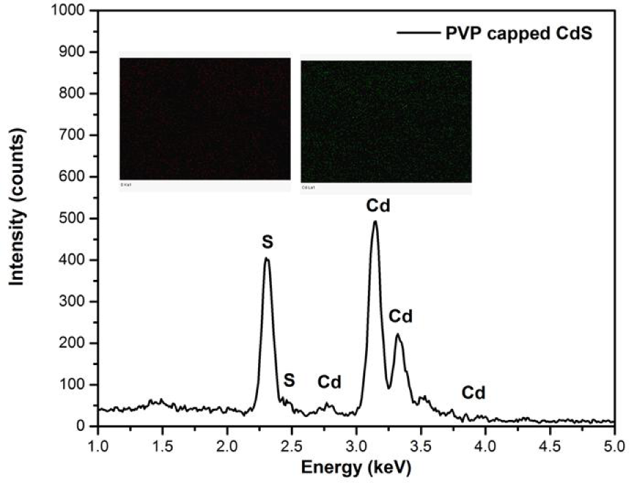
EDX and elemental mapping of PVP capped CdS nanoparticles
Fig.4 (a-c) shows the TEM images indicating that the PVP capped CdS nanoparticles have monodispersed spherical crystallites. By the statistical means on the HRTEM image in the inset in Fig. 4c clearly revealed the synthesised CdS nanoparticles have particle size of ∼3 nm, which is corroborated with the XRD results. The nanoparticles are clearly well identified and no effective aggregation of bulk particles is formed, indicating effective capping of PVP on the nanoparticle surfaces. The reason for the appearance of aggregates of CdS nanoparticles possibly is static attraction of their surface groups. From the TEM images, it is observed that PVP plays an important role in enhancing the monodisperse property of CdS nanoparticles. In this process PVP, lowers the surface energy of the nanoparticles, and hence the PVP capped CdS nanoparticles shows excellent monodispersive property [16]. PVP can modify the Ostwald ripening kinetics in such a way that the growth rate decreases with the size of the CdS nanoparticles and effectively narrow the size distribution [17]. Lattice fringe pattern and size of CdS nanocrystallites are clearly reveal from the inset of Fig. 4(c). The high-resolution image also (Fig. 4(d)) confirms the synthesised particles are crystalline in nature.
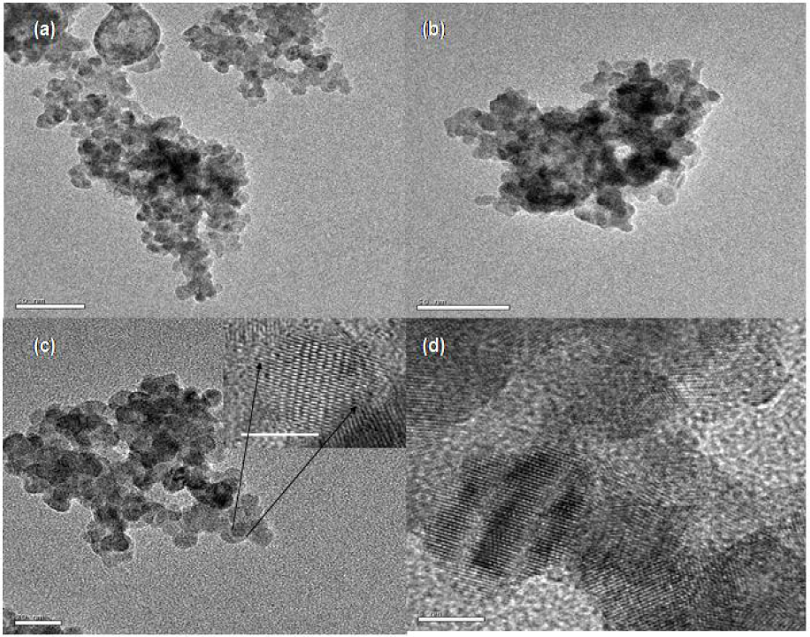
TEM and HRTEM (inset) micrographs of PVP capped CdS nanocrystallites. The length bar of (a) and (b) is 50 nm and for (c) is 20 nm. The scale of HRTEM in inset Fig. (c) is 2 nm and in (d) is 5 nm.
3.2 Optical analysis
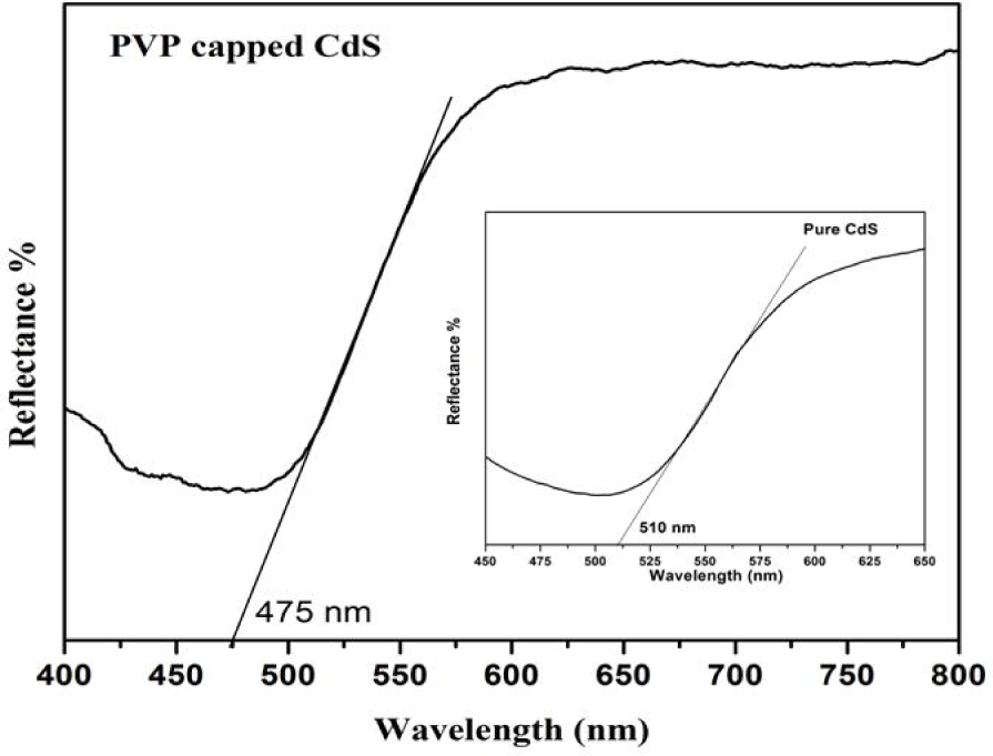
In order to determine the band gap of PVP capped CdS nanoparticles, reflectance UV spectrum were recorded as shown in Fig.5. The estimated band gap value for PVP capped CdS is 2.61 eV corresponding to the absorption edge at 475 nm. The inset in Fig.5 shows the UV-reflectance spectrum of pure CdS with an absorption edge at 510 nm synthesised without capping agent, which is blue shifted compared with the absorption edge of the bulk CdS (520 nm). Because the binding energy of the exciton increases with decreasing size due to the increasing columbic overlap enforced by spatial localization of the wave functions, as the shift in the band gap with size dominates the spectral changes [12].
DRS - UV spectrum of PVP capped CdS nanoparticles (inset shows DRS - UV spectrum of pure CdS)
This blue shift in the optical absorption edge indicates the formation of CdS particles in the nanometer regime. Similar results observed for CdSe nanoparticles in the PVP-PVA matrix with blue shift in the absorption spectrum [18]. It has been reported that PVP not only controls the particle size but also reduces the wider distribution of the nanoparticles in the matrix.
Fig. 6 shows the FTIR spectrum of PVP capped CdS nanoparticles. The broad absorption band centered at 3437 cm−1 is attributed to O-H stretching mode of H2O absorbed on the surface of the product. The most striking evidence from FTIR spectrum of the PVP stabilized CdS is the broad peak between 1250 and 1650 cm−1 which corresponds to C-N stretching motion and C=O stretching motion of monomer for PVP, respectively [19–20]. The narrow absorption peak centered at 1409 cm−1 and 2876 cm−1 occurred in Fig. 6 is ascribed to the C-H bonding due to the presence of PVP [21]. This may be due to the formation of coordinate bond between the nitrogen atom of the PVP and the Cd2+ ions, similar to the previous reports [22–23]. FTIR spectroscopy is well suited for quantitative determination of sulfur oxygen anions in the strong IR-absorbing aqueous medium. The relatively intense S-O stretching absorption bands in the 750 to 1350 cm−1 region of the IR spectrum shows the presence of Sulfur-oxygen compounds. Results of the FTIR spectroscopy confirm that the surface of the synthesised CdS nanoparticles is modified with PVP.

FTIR spectrum of PVP capped CdS nanoparticles
Fig. 7 shows the photoluminescence spectrum of PVP capped CdS nanoparticles. The first peak is assigned due to the band gap transitions, while the second one is due to sulfur vacancy in the CdS nanoparticles. At nanometric sizes, quantum confinement effects come into play and affect most notably the electronic properties [24].

Room temperature photoluminescence spectrum of PVP capped CdS nanoparticles (inset shows PL spectrum for uncapped CdS)
Therefore, the perceptible attention has been paid to prevent the agglomeration of CdS particles in order to improve their photoluminescence properties. A room temperature photoluminescence measurement shows that the sample emits a stable bluish light and broad luminescence at 396 nm. This strong emission peak at 396 nm, assigned to the electron-hole recombination of CdS [25], while the other lower emission peak might be assigned to the surface trap induced emission owing to the PVP capping. The enhanced luminescence properties of the capped CdS nanoparticles are due to the surface modification by PVP molecule, with the effect of minimizing surface defects and enhance the possibility of electron-hole recombination as reported earlier [26]. This result clearly justifies that the PVP as the capping agent can significantly enhanced the PL intensity for CdS nanoparticles.
The green luminescence peak position shifts to the red from 504 nm (non-PVP capped sample) to 520 nm due to PVP capping. These observations suggest that the green emission originates mainly from the deep surface traps, which can be removed via surface passivation by PVP. The PVP-induced red shift of the green emission peak may be explained by the inhomogeneity of the surface traps, because the PVP molecules remove the shallower surface traps more effectively, leaving behind the deeper surface traps or inner defects. The ability of CdS nanocrystallites to emit photoluminescence is enhanced after nanocrystallites are stabilized using polyvinyl pyrrolidone and maintaining high fluorescent intensity compared to uncapped CdS nanocrystallites.
Raman spectra of the pure CdS and PVP capped CdS nanoparticles excited at 514.5 nm are depicted in Fig.8. The scattering peaks at 296, 596 and 893 cm−1 correspond to first order (1-LO), second order (2-LO) and third order (3-LO) lower frequency longitudinal-optical (LO) phonon modes of zinc-blende phase cadmium sulfide respectively. Moreover, the relatively sharp and symmetric profile [27] of the peaks of our sample suggests that synthesised nanoparticles are highly crystalline and relatively free of impurities. We observed the LO peaks of PVP capped CdS nanoparticles shifted towards lower frequency. With the reduction of size, the surface/volume ratio increases and the phonon confinement induced by the size reduction plays an important role in determining the physical properties of semiconductor nanocrystals, resulting in the higher energetic state of their surface atoms [28–29].
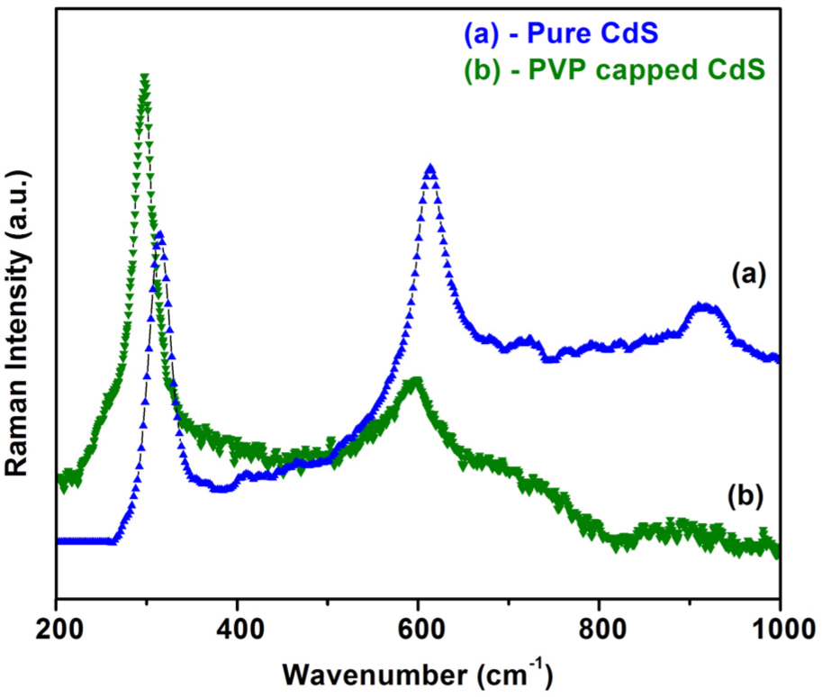
Micro-Raman spectra of (a) pure CdS and (b) PVP capped CdS nanoparticles
The observed shift in the phonon peaks toward lower frequency would be expected from bulk, likely due to effects of small size and high surface area [30]. Yang et al also developed a model, stated that the observed Raman red shift is caused by the combination of the effects of size-induced phonon confinement and surface relaxation [31].
3.4 Mechanism of PVP capping
In this investigation polyvinyl pyrrolidone (PVP), a water soluble polymer, was used as capping molecules and also to stabilize the CdS nanoparticles. The pyrrolidone part (hydrophilic) acted as the head group while the polyvinyl part (hydrophobic) acted as the tail group. The role of the PVP is twofold: (a) either it controls the growth of the particles by forming passivation layers around the CdS core via coordination bond formation between the nitrogen atom of the PVP and Cd2+ ion, and/or (b) it prevents agglomeration by steric effect due to the repulsive force acting among the polyvinyl groups (tail part). Therefore, the PVP encapsulation creates a restricted environment around the CdS nanocrystals [22].
Addition of large amount of PVP on the particle surface may cause the attraction among their polymeric chains due to the osmotic pressure. This phenomenon is known as “depletion flocculation”, which causes destabilization [32]. Competitive kinetics between the binding of cadmium to the carboxylate functional groups in a growth termination step or to the sulfide ions in initiation and propagation steps leads to the growth of CdS cluster whose surface is capped and whose dimensions are trapped in the nanometer size regime, as determined by the relative rates of the propagation and termination steps [33].
4. Conclusion
Monodispersive cubic zinc blende phase of spherical CdS nanoparticles have been synthesised by chemical coprecipitation method using PVP as a capping agent. The average crystallite size of ∼3 nm was estimated by XRD and was confirmed with HRTEM. The blue shift to 475 nm can be compared with the bandgap of the characteristic absorption of uncapped CdS, which was probably due to the size quantization effect. FTIR and Raman spectroscopic measurements have revealed the presence of PVP molecule on the surface and the observed red shift in the optical phonon modes of CdS. The results are attributed to the effect of reducing surface defects and enhancing the possibility of electron-hole recombination of the CdS nanoparticles by capping the surface with the PVP molecules. The results infer that PVP is an effective polymer to improve the luminescence properties of CdS nanoparticles.
Footnotes
5. Acknowledgments
The author thanks Dr. K.V.R Murthy, Applied Physics Department, Faculty of Technology and Engineering, M. S. University of Baroda, India for the help in recording the PL measurement and his support to this work.