
Abstract
Purpose
The aim of the present in vitro study was to evaluate the leakage observed in external hexagon (EH) and cone Morse (CM) tapered implant-abutment connections, using toluidine blue.
Methods
A total of 60 implants, 30 with a screw-retained EH abutment and 30 with a CM taper internal connection, were used. Toluidine blue was placed into the deepest portion of the internal compartment of the 2 different implant systems, and cyclic loading was applied for each group as follows: 10 samples underwent 1 × 10⁶ loading cycles, 10 samples underwent 3 × 10⁶ cyclic loading and the least 10 samples underwent 6 × 10⁶ cyclic loading.
Results
No significant differences between the EH and CM groups were detected when the lowest loading cycles were applied (p = 0.2624), while differences were found when the samples were loaded with 3 x 10⁶ and 6 x 10⁶ cycles (p = 0.00124), with significantly lower toluidine leakage in CM group.
Conclusions
In conclusion, the results of the present in vitro study demonstrated that flow of the toluidine blue to the external portion of the implant-abutment assembly occurred in both types of implant-abutment connections, with very different percentages. Indeed, the CM taper internal connection seems to be more resistant to the leakage of dyes when compared with EH connections.
Keywords
Introduction
Peri-implant inflammatory reaction seems to be mainly related to bacterial leakage at the implant-abutment junction (IAJ) (1). After the implant and abutment connection, the formation of cavities has been described; these cavities or gaps can represent a reservoir for bacteria (2), and furthermore, they may determine an unfavorable stress distribution on the different connection components – on the implant and on the crestal peri-implant bone (3). The precision of fit between the different components (implant and abutment), the various degrees of micromovement between the same components and the torque forces used for the implant-abutment connection determine the different degrees and the varying amounts of the movement of bacteria (from the inside to the outside and vice versa) in the different implant systems (4). The action of bacterial reservoir in the implant gaps and cavities can produce a resorption of the peri-implant bone and an inflammatory infiltrate of the soft tissues (2, 5-6-7).
Bacterial penetration has been found to occur in static conditions, and seems to be increased when the assemblies are loaded. Loading forces on the prosthetic restoration under function may cause bending of the components or movements inside the implant system, with the formation of a larger gap, determining a “pump” effect that probably increases the movement of the microorganisms (4). An inflammatory cell infiltrate (infiltrated connective tissue [ICT]) has been reported, in several in vivo experimental studies, to be present at the level of the IAJ (5, 6, 8). Inflammatory events at the IAJ and peri-implant crestal bone loss seem to be closely related (7, 9, 10). An apical placement of the IAJ, advocated, from a clinical point of view, mainly for esthetic reasons, seems to be likely to produce the largest degree of bone loss (11, 12). The IAJ microgap probably represents an active infection site, with an inflammatory response from the host (10). Many techniques have been used, in vitro, to evaluate the sealing capabilities in different implant-abutment connections – e.g., bacteria, bacterial toxins, dyes (toluidine blue and gentian violet), gas, saliva etc. Even if most of the studies reported in the literature have been done with the use of different types of bacteria, there are advantages in using dyes – i.e., a similar size of the particles, easy use, capability to quantify the results (13).
The aim of the present in vitro study was an evaluation of the leakage observed, in external hexagon and cone Morse tapered implant-abutment connections, using toluidine blue.
Materials and methods
A total of 60, 4 × 13 mm, implants were used in this in vitro study, 30 with a screw-retained external hexagon abutment and 30 with a cone Morse taper internal connection (Universal II HI and CM, respectively; Implacil, De Bortoli, Sao Paulo, Brasil). First, the samples were divided into 2 groups by the type of connection: the external hexagon connection (group EH) (30 implants) and the cone Morse connection (group CM) (30 implants). Each group was further subdivided into group A (10 samples), group B (10 samples) and group C (10 samples) according to the different loading cycles: group A underwent 1 × 10⁶ loading cycles; group B, 3 × 10⁶ cyclic loading; and group C, 6 × 10⁶ cyclic loading. The loading force was applied to the abutment. A customized jig was designed to hold the implant, and the distance was 3 mm from the implant platform to the exposed position of the implant. The distance of 3 mm was chosen to represent the worst case in bone retraction. The load was applied on the abutment at 30°C, and the distance was 11 mm from the center of the hemisphere to the exposed position of the implant. Then, each sample was loaded in a cyclic loading mode with a Lloyd 30K universal testing machine (Lloyd Instruments Ltd, Segensworth, UK), which controls the 20-300 N/cm cyclic loading in an Hsine shape at 4 Hz (ISO/DIS 14801: Dental implants-Dynamic continuous fatigue test, 2003). An electronically controlled automated pipette was then utilized to place 0.7 µL of toluidine blue into the deepest portion of the internal compartment of the 2 different implant systems (Fig. 1A, B). This procedure was facilitated due to the small dimension of the pipette tip, which was easily inserted through the whole depth of all implant systems internal threads. Following the color marker placement inside the implant connection, the abutments were connected to the implants according to the manufacturer's recommendations. The connected implant abutments were placed into 15-mL vials previously filled with 3 mL of distilled water.
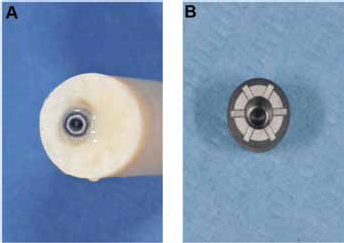
Toluidine blue dye was placed into the deepest portion of the internal compartment of the external hexagon
Statistical evaluation
The differences in the presence or absence of toluidine blue were statistically analyzed using the Mann-Whitney U test. Statistically significant differences were set at a p value <0.05.
Results
Group EH (external hexagon connection)
In the samples of group A, toluidine blue was found in 1 of 10 samples. In group B, toluidine blue was found in 3 of 10 samples. In group C, toluidine blue was found in 6 of 10 samples (Fig. 2A).
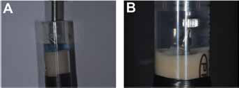
The connected implant abutments were placed into vials previously filled with distilled water.
Group CM (cone Morse connection)
In groups A and B, toluidine blue was absent in all the samples (Fig. 2B). In group C, toluidine blue was found in 1 of 10 samples.
Statistical Analysis
Statistical analysis showed no significant differences between the EH and CM groups when the lowest loading cycles (group A) were applied (p = 0.2624), while differences were detected between groups B and C (p = 0.00124), with significantly lower toluidine leakage in the CM group.
Discussion
The problem of a microgap between implant and abutment is biological and mechanical. The biological problem relates to the presence of bacteria that, in vivo, can produce a bacterial reservoir that, on turn, interferes with the long-term health of the peri-implant tissues and with the long-term prognosis of the implant (14). The mechanical problem relates to micromovement and possible loosening or fracturing of screw-retained abutments (14). The precise mechanism responsible for the crestal bone remodeling in 2-piece implants is not known (15). The existence of bacterial leakage, both at the junction between the abutment and the implant, and along the abutment screw has been reported (16). This crestal bone resorption has not been observed around sleeping implants, where both exposure to microbial colonization and loading were absent (16). It has been shown that inflammation results if the abutment loosens on the implants placed in a submerged approach, with a possible fistula formation (17). There is a physiological reaction to the presence of an interface; the reason for this reaction is unknown, but may be related to the presence of bacteria contamination or micromovements of the interface (9). In a histological study of 2 human screw-retained implants, retrieved at autopsy, a gap was present between the implant and the healing screw, and this space was filled by bacteria and calculus (18). Bacteria were also present in the most apical portions of the hollow part of the implants, and an inflammatory infiltrate was present in the connective peri-implant tissues (18). The presence of the inflammatory infiltrate (ICT) confirmed, in an in vivo human study, the data reported in animal experimental studies. In a retrospective microscopical study of human implants with a screwed-retained abutment, retrieved after a long period of clinical service, bacteria were often found in the microgaps between implant and abutment and in the internal portion of the implants (14). In a retrospective histological study in monkeys, it was found that no inflammatory infiltrate was present when the implants had been inserted with the microgap above the alveolar crest level, while, on the contrary, many inflammatory cells were present in the area of the IAJ and inside the gap, with many osteoclasts resorbing bone, in implants that had been placed at the level or below the alveolar crest (8). In conclusion, the results of the present in vitro study demonstrated that flow of the toluidine blue to the external portion of the implant-abutment assembly occurred in both types of implant-abutment connections, even if with very different percentages. In the cone Morse assemblies, a very low occurrence of leakage was found only in the samples that underwent 6 x 10⁶ cycles of loading, while no leakage was present in the samples of the groups undergoing 1 x 10⁶ and 3 x 10⁶ loading cycles. On the other hand, the external hexagon specimens showed a leakage in specimens of the 3 groups (A, B and C). There was an increase of the percentage of leakage with an increase of the loading cycles. When making a statistical evaluation between the samples of the 2 groups, a statistically significant difference was found in the specimens undergoing 3 x 10⁶ and 6 x 10⁶ loading cycles.
In conclusion, the cone Morse taper internal connection seems to be able to resist more of the leakage of dyes, when compared with external hexagon connections.
Footnotes
Financial support: This work was partially supported by the Ministry of Education, University, and Research (M.I.U.R.), Rome, Italy.
Conflict of interest: The authors state they have no conflict of interest.