
Abstract
For both lung cancer patients and clinical physicians, tumor biomarkers for more efficient early diagnosis and prediction of prognosis are always wanted. Biomarkers in circulating serum, including microRNAs (miRNAs) and extracellular vesicles, hold the greatest possibilities to partially substitute for tissue biopsy. In this systematic review, studies on circulating or tissue miRNAs and extracellular vesicles as potential biomarkers for lung cancer patients were reviewed and are discussed. Furthermore, the target genes of the miRNAs indicated were identified through the miRTarBase, while the relevant biological processes and pathways of miRNAs in lung cancer were analyzed through MiRNA Enrichment Analysis and Annotation (MiEAA). In conclusion, circulating or tissue miRNAs and extracellular vesicles provide us with a window to explore strategies for diagnosing and assessing prognosis and treatment in lung cancer patients.
Introduction
Lung cancer is one of the most common and serious types of cancer. As the leading cause of cancer-related deaths in males and second most common cause in females (1), lung cancer contributes greatly to the global disease burden. Early diagnosis of lung cancer improves the overall survival rate of patients significantly. However, the average 5-year survival rate of advanced lung cancer is as low as 3.6%. The difficulties in early diagnosis are due to a lack of signs or symptoms in the early stages (2). Therefore, identifying new molecular markers for early detection is urgently needed.
MicroRNAs (miRNAs) and extracellular vesicles are deeply involved in cancer pathogenesis. The profiles of miRNA levels are believed to be correlated with pathological conditions. MiRNA and extracellular vesicles derived from cancer cells may be packaged in exosomes and microparticles. and provide information regarding tumor progression, reflect the status of pathological conditions and serve as biomarker candidates for diagnosis and predicting prognosis, as well as for assessing therapy response and side effects.
Adequate sensitivity and specificity in early diagnosis of lung cancer was shown to be achieved by a combination of miR-21, miR-143, miR-155, miR-210 and miR-372 in a previous study (3). The role of serum miR-25, miR-145 and miR-210 in predicting the prognosis of advanced non-small cell lung cancer (NSCLC) patients was also suggested by reports in the literature (4). NSCLC patients with higher levels of miR-4782-3p had more favorable outcomes (5). Circulating miR-29a and miR-150 were suggested to be related to thoracic radiation therapy for NSCLC patients (6). Therefore, a systematic review of these studies would be very useful to explore the implications of miRNAs as biomarkers in lung cancer patients.
Additionally, circulating extracellular vesicles (EVs), including exosomes and microparticles, are small membrane vesicles budding from several kinds of cells. Recent studies showed that, in advanced NSCLC patients, the circulating level of endothelial-derived microparticles (EMPs) was correlated with 1-year mortality (7). Here, the published researches were systemically reviewed to discuss the role of miRNAs and EVs as promising biomarkers in lung cancer patients.
Material and methods
Search strategy and data extraction
The MEDLINE database was searched using Pubmed for published studies from 1988 until March 24, 2017. The search phrases were (((((“patients”[MeSH Terms] OR “patients”[All Fields]) OR (“patients”[MeSH Terms] OR “patients”[All Fields] OR “patient”[All Fields])) AND ((“lung”[MeSH Terms] OR “lung”[All Fields]) OR (“lung”[MeSH Terms] OR “lung”[All Fields] OR “pulmonary”[All Fields]))) AND ((“neoplasms”[MeSH Terms] OR “neoplasms”[All Fields] OR “cancer”[All Fields]) OR (“tumour”[All Fields] OR “neoplasms”[MeSH Terms] OR “neoplasms”[All Fields] OR “tumor”[All Fields]) OR (“tumour”[All Fields] OR “neoplasms”[MeSH Terms] OR “neoplasms”[All Fields] OR “tumor”[All Fields]) OR (“neoplasms”[MeSH Terms] OR “neoplasms”[All Fields]) OR malignant[All Fields] OR (“carcinoma”[MeSH Terms] OR “carcinoma”[All Fields]))) AND ((“micrornas”[MeSH Terms] OR “micrornas”[All Fields]) OR (“micrornas”[MeSH Terms] OR “micrornas”[All Fields] OR “microrna”[All Fields]) OR (“exosomes”[MeSH Terms] OR “exosomes”[All Fields] OR “exosome”[All Fields]) OR (“exosomes”[MeSH Terms] OR “exosomes”[All Fields]) OR (“extracellular vesicles”[MeSH Terms] OR (“extracellular”[All Fields] AND “vesicles”[All Fields]) OR “extracellular vesicles”[All Fields] OR (“extracellular”[All Fields] AND “vesicle”[All Fields]) OR “extracellular vesicle”[All Fields]))) NOT review[Publication Type].
Exclusion criteria
The exclusion criteria were (i) not a human study, (ii) not a lung cancer study, (iii) not an miRNA study, (iv) not about lung cancer biomarkers, (v) a review article, (vi) article not in English and (vii) article not accessible.
Target gene, biological process and pathway analysis
The target genes of miRNAs were determined using the miRTarBase database (http://mirtarbase.mbc.nctu.edu.tw/, accessed 2 October 2017). The MiRNA Enrichment Analysis and Annotation database (https://ccb-compute2.cs.uni-saarland.de/mieaa_tool/user_input/, accessed 2 October 2017) was used to analyze the relevant biological processes and pathways of miRNAs.
Results
A total of 1091 published studies were retrieved by the search strategy above. A total of 571 articles were excluded due to the exclusion criteria. Five hundred and twenty articles related to miRNAs or EVs as lung cancer biomarkers were enrolled. Further, studies that discussed miRNA single nucleotide polymorphisms (SNPs) or had negative results were excluded. both Using these criteria, 455 high-quality studies were finally included in this analysis (Fig. 1).
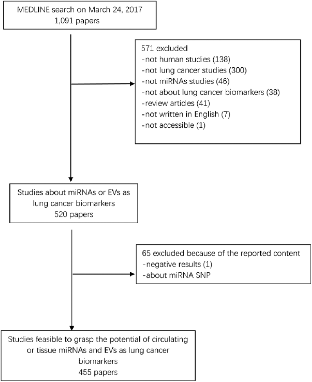
Flow chart of this systematic review. A total of 1,091 papers were identified via the initial search, and 455 papers were finally included. EVs = extracellular vesicles; miRNA = microRNA; SNP = single nucleotide polymorphism.
From the result shown in Figure 2A, it can be seen that the publications increased rapidly year by year from 2008. As for the pathological classification of lung cancer investigated, NSCLC, including adenocarcinoma (AD) and squamous cell carcinoma (SCC), was discussed very fully. In addition, small cell lung cancer (SCLC) was also discussed in 10 papers. Specifically, there were 3 papers about metastatic lung cancer, 1 paper about smoking-associated lung cancer and 1 paper about AD-associated pleural effusion (Fig. 2B). China is the leading source of publications (59.6%), while other countries have also contributed greatly to the studies reported (Fig. 2C). Among these 455 publications, 246 papers (54.1%) assessed efficacy of diagnosis, 191 (42.0%) explored the predictivity of prognosis, 63 (13.8%) evaluated both diagnosis and prognosis prediction, 23 (5.1%) evaluated prediction of chemotherapeutic response and radiochemotherapy side effects, 20 (4.4%) evaluated predicted metastatic outcomes and 3 (0.7%) evaluated prediction of tumor relapses (Fig. 2D).
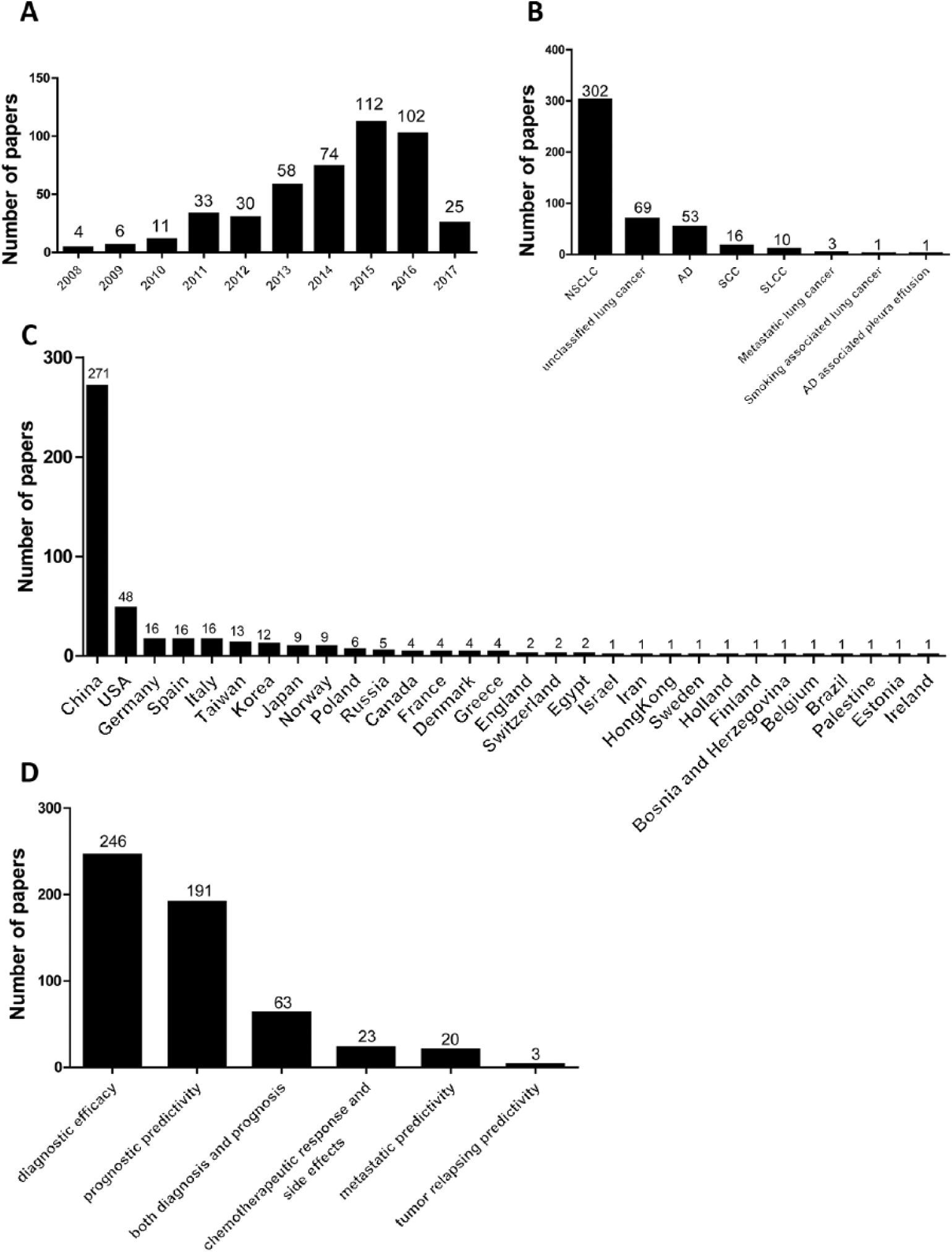
Basic characteristics of the publications included. Numbers of papers year (
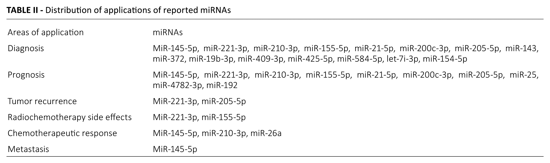
Of all of the 455 papers, there were 438 papers (96.3%) related to miRNAs. In the present study, we listed the top 10 miRNAs discussed, as well as their functions as lung cancer biomarkers, and the top 3 target genes (Tab. I). Different miRNAs and their biological functions are listed in Table II. Several high-quality studies are presented as examples to show these miRNAs as promising biomarkers in patients with lung carcinoma.
The 10 most frequently documented miRNAs in lung cancer
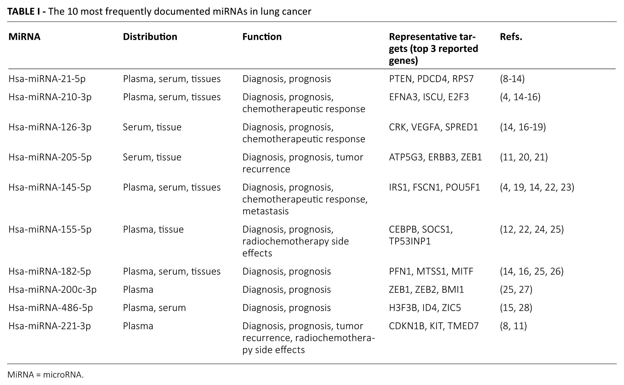
MiRNA = microRNA.
Distribution of applications of reported miRNAs
Circulating miRNAs
A 4-phase study, which included 141 lung adenocarcinoma (LA) patients and 124 healthy controls, conducted by the Cancer Center of Nanjing Medical University (8), screened potential miRNAs for early LA detection, by testing concentrations of plasma miRNAs. Furthermore, the identified miRNAs in lung cancer tissue samples and exosomal miRNAs were confirmed. Up-regulation of miR-19b-3p, miR-21-5p, miR-221-3p, miR-409-3p, miR-425-5p and miR-584-5p was found in LA patients. These 6 miRNAs discriminated LA patients from healthy controls with an area under the curve (AUC) of >0.72. Specifically, dramatically increased expression levels of miR-584-5p were detected in lung cancer tissues, while increased levels of miR-19-3p, miR-21-5p, miR-409-3p and miR-425-5p were detected in LA patients’ plasma, and increased levels of miR-19-3p, miR-21-5p and miR-221-3p were observed in plasma exosomes in LA patients. These results may provide valuable suggestions for approaches to achieving early detection of LA to a certain extent, especially for Asian patients.
Cigarette smoking is the major risk factor for lung carcinoma. In research performed by Huang et al (29), 10 patients with lung cancer, 10 smokers and 10 nonsmokers were enrolled, and their sera miRNA expression was detected and compared. A study with larger samples was performed to confirm the changed miRNAs. Compared with nonsmokers, patients with lung cancer and smokers had higher sera levels of let-7i-3p and miR-154-5p. Therefore, we can conclude that there is a relationship between circulating let-7i-3p (AUC = 0.892) and miR-154-5p (AUC = 0.957) and smoking, as well as smoking-related lung cancer. This research revealed a relationship between circulating miRNAs and risk of cigarette-associated LAs and provides evidence for let-7i-3p and miR-154-5p as promising biomarkers in cigarette-associated LAs.
Radiochemotherapy is the most important part of lung cancer treatment, especially for patients with advanced stages of the disease. However, radiochemotherapy side effects remain a major concern for clinicians. A study performed by the Duke Cancer Institute assessed the relationship between severe radiation-induced esophageal toxicity (RIET) and circulating miRNAs (miR-155, miR-221 and miR-21) in 101 NSCLC patients (30). The results indicated that in the initial 1-2 weeks of chemoradiation therapy, patients with higher expressions of miR-155 and miR-221 had a higher risk of suffering severe RIET (p<0.05, for both miR-155 and miR-221). In addition, patients with a several fold alteration of miR-221 were more likely to suffer from severe RIET (p<0.05).
Tissue miRNAs
As SCLC has a shorter doubling time and faster growth rate, more easily metastasizes and has a poorer prognosis, it is considered to be an extremely malignant kind of cancer. Besides, there are few biomarkers for detecting SCLC and predicting its prognosis. Mancuso et al (27) detected the expression levels of miR-192, miR-200c and miR-205 in 50 SCLC patients, with 25 primary tumors and 25 metastases. They showed that patients with lower expression of miR-192, miR-200c and miR-205 had a more favorable prognosis.
A study conducted by Nanjing University investigated the expression levels of miR-26a and its target gene, enhancer of zeste homolog 2, in docetaxel-sensitive and docetaxel-insensitive advanced LA patients (31). Lower expression of miR-26a was found in docetaxel-sensitive patients compared with docetaxel-insensitive patients. Investigations into the mechanisms suggested that miR-26a could inhibit the proliferation and epithelial-to-mesenchymal transition (EMT), and also increase apoptosis of docetaxel-resistant LA cells.
Biological process and pathway analysis
The genes that harmonize biological pathways often require synergistic effects to accomplish unified biological functions. Biological process and pathway analysis can increase an understanding of miRNA function. As shown in Figure 3, 19 kinds of biological processes were found among 207 miRNAs, including platelet-derived growth factor receptor binding, positive regulation of cell adhesion, receptor binding, cell aging, positive regulation of endothelial cell proliferation, chromatin, induction of positive chemotaxis proteinaceous extracellular matrix, coated pit, mesenchymal cell development, oligodendrocyte differentiation, positive regulation of leukocyte migration, nuclear membrane, positive regulation of cell growth, metanephros development, negative regulation of ossification, membrane raft, positive regulation of gene expression and regulation of cell proliferation. In Figure 4, the top 11 pathways are listed, including apoptosis, extracellular matrix (ECM) receptor interaction, P00052 TGF beta signaling pathway, p38 MAPK signaling pathway, hsa04350 TGF beta signaling pathway, ubiquitin mediated proteolysis, NOD-like receptor signaling pathway, cytokine–cytokine receptor interaction, endocytosis, cell adhesion molecules and Wnt signaling pathway.
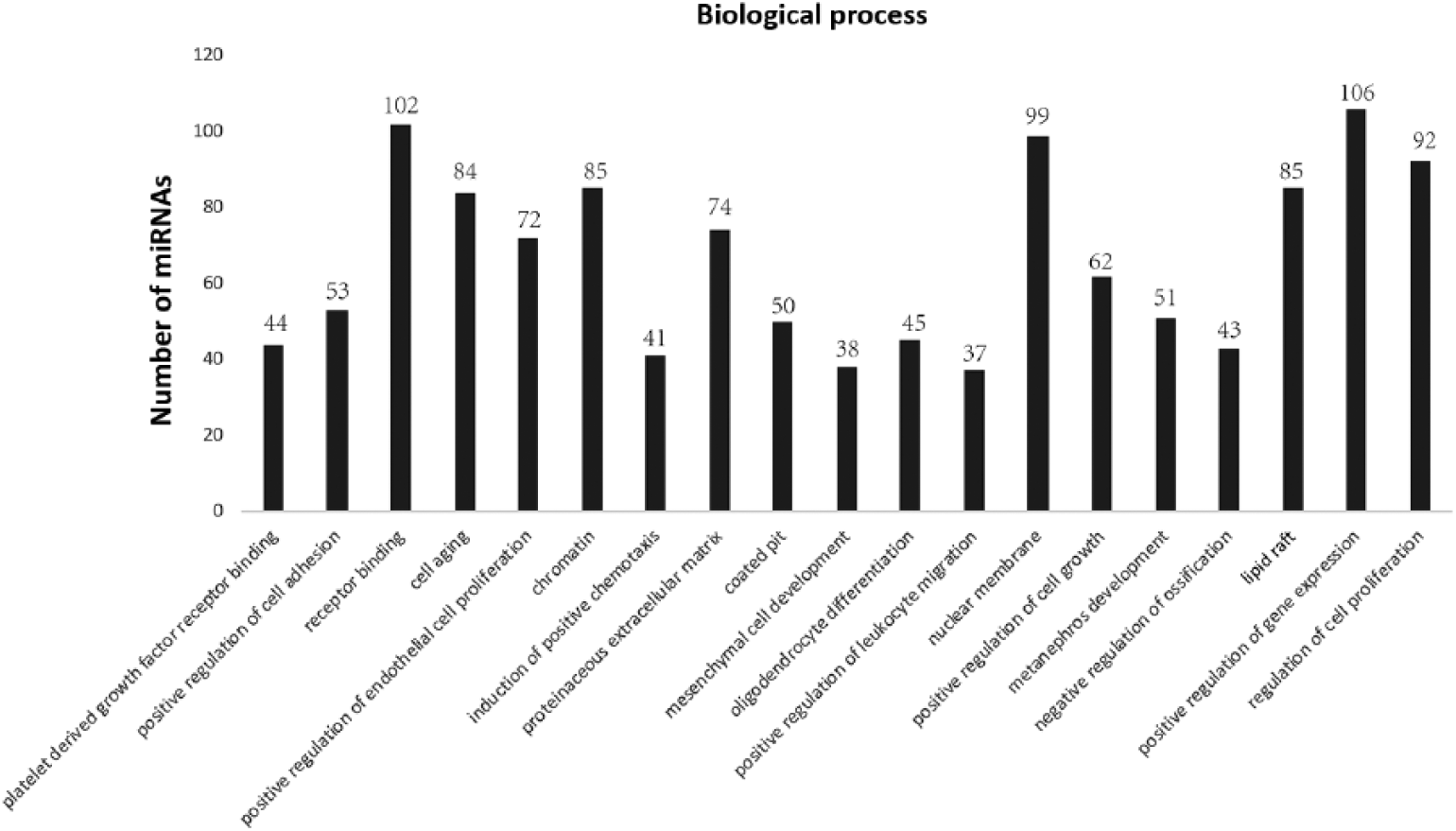
Biological process analysis of indicated microRNA (miRNAs) in this review. Through biological process analysis via MiRNA Enrichment Analysis and Annotation (MiEAA), 19 major biological processes for the indicated miRNAs in lung cancer were identified.
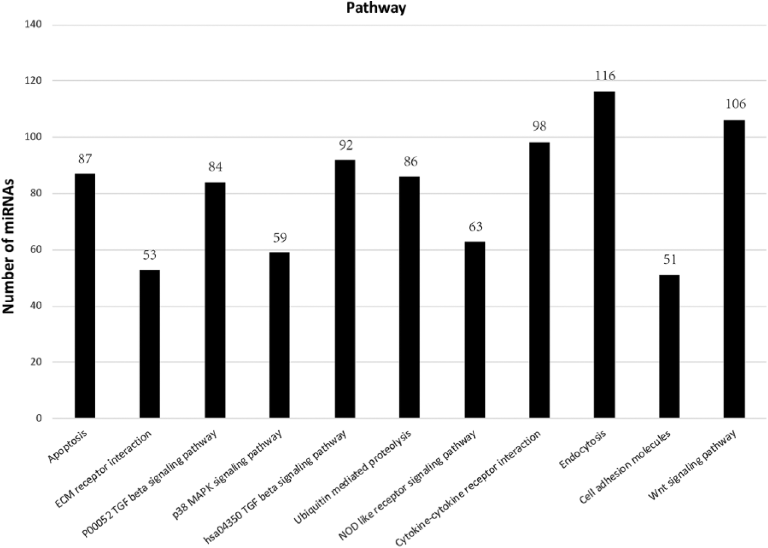
Pathway analysis of indicated microRNA (miRNAs) in this review. Through pathway analysis via MiRNA Enrichment Analysis and Annotation (MiEAA), 11 major pathways for the indicated miRNAs in lung cancer were identified. ECM = extracellular matrix; TGF = transforming growth factor.
EV-associated proteins
In our present systematic review, there were 17 papers (3.7%) on EV-associated proteins and microparticles. Besse et al (32) performed a phase II clinical trial to determine the effects of dendritic cell–derived exosomes with IFN-γ (IFN-γ-Dex) as maintenance immunotherapy for late-stage NSCLC patients who received chemotherapy. They showed that IFN-γ-Dex activated NK cell functions and correlated with prolonged progression-free survival in patients.
Exosomal proteins as diagnostic and prognostic biomarkers in NSCLC patients were also confirmed by Sandfeld-Paulsen et al (33, 34). They measured exosomal protein profiles in 431 NSCLC patients and 150 healthy subjects to determine any potential role of exosomal proteins for diagnosing lung cancer (33). CD151, CD171 and tetraspanin 8 were proven to be the strongest markers for discriminating between NSCLC patients and healthy subjects. Moreover, multimarker models were more suitable for diagnosis of patients with AD only (AUC = 0.76). At the same time, they evaluated exosomes as prognostic biomarkers by protein phenotyping in 276 NSCLC patients (34). A total of 49 proteins, which were found to be expressed in the membranes of exosomes, were investigated in their study. Finally, after multiple testing and adjustment, they suggested that higher levels of New York esophageal squamous cell carcinoma-1 (NY-ESO-1) were related to a worse prognosis.
Levels of circulating platelet-derived microparticles (PDMPs) and endothelial-derived microparticles (EDMPs) as promising prognostic markers for patients with lung cancer were also explored by researchers (7, 35). First, they detected the circulating levels of platelet-derived activated microparticles (PDAc-MPs), platelet-derived apoptotic MPs (PDAp-MPs), endothelial-derived activated MPs (EDAc-MPs) and endothelial-derived apoptotic MPs (EDAp-MPs) in 130 lung cancer patients (35). They found that the levels of these 4 MPs were significantly lower in lung cancer patients than in healthy controls (all p<0.05). In addition, lower levels of PDAc-MPs were found in patients with early-stage of lung cancer compared with patients with advanced-stage lung cancer (p = 0.031), while levels of EDAp-MPs were related to the different cell types, such as SCC, AD and SCLC (p<0.05). Furthermore, they indicated that lower levels of circulating EDAc-MPs were found in patients with more than 1 year of survival compared with patients with less than 1 year of survival (p = 0.006). Further multivariate analysis indicated that circulating levels of EDAc-MPs along with brain metastasis and male sex predicted 1-year mortality significantly and independently (all p<0.05) (7).
Conclusions and expectations
In the present systematic review, a total of 455 papers regarding the potential role of miRNAs and EVs as biomarkers for lung cancer diagnosis and prognosis prediction were identified. We can conclude that circulating or tissue miRNAs and EVs provide us with a window to explore strategies for diagnosing and assessing prognosis and treatment in lung cancer patients. However, there are still several limitations to the current studies. A considerable proportion of diagnostic and prognostic studies failed to achieve a high degree of accuracy (AUC>0.8). In addition, many studies were restricted to only detecting the expression levels of related miRNAs and EV-associated proteins. And some functional studies did not explore deeply into the molecular pathways of miRNAs in lung cancer either. Although there is a long way to go to enable lung cancer patients to really benefit, miRNA and EVs screening will bring us important improvements in lung cancer practice in the near future.
Footnotes
Disclosures
Financial support: No grants or funding have been received for this study.
Conflict of interest: None of the authors has any financial interest related to this study to disclose.