
Abstract
Fluorine-19 (19F)-based contrast agents for magnetic resonance imaging stand to revolutionize imaging-based research and clinical trials in several fields of medical intervention. First, their use in characterizing in vivo cell behavior may help bring cellular therapy closer to clinical acceptance. Second, their use in lung imaging provides novel noninvasive interrogation of the ventilated airspaces without the need for complicated, hard-to-distribute hardware. This article reviews the current state of 19F-based cell tracking and lung imaging using magnetic resonance imaging and describes the link between the methods across these fields and how they may mutually benefit from solutions to mutual problems encountered when imaging 19F-containing compounds, as well as hardware and software advancements.
Introduction
Fluorine-19 (
19
F) has the potential to revolutionize imaging-based cell tracking, lung imaging, and drug development by enabling alternate routes for clinical translation of these techniques to patients.
19
F is a versatile and nuclear magnetic resonance (NMR) active nucleus. Observations were originally attempted in 1942 using NMR
1
and later with magnetic resonance imaging (MRI) in the 1970s,
2
when numerous applications were surmised.
19
F T1 relaxation rates were first used in the early 1990s to determine the intracellular partial pressure of oxygen, but direct fluorine imaging still proved elusive.
3
Technology has caught up with the predictions of the past and MRI contrast agents containing
19
F are now at the forefront of imaging research in a variety of biomedical fields such as cell tracking, lung imaging and drug development. Figure 1 clearly demonstrates an increase in the number of publications for
19
F-based MRI cell tracking within the past decade. From an MRI point of view,
19
F is part of the family of nonproton nuclei (or X-nuclei). It is 100% naturally abundant and has a spin of 1/2, a gyromagnetic ratio of appreciable size (94%, the largest of X-nuclei) compared with protons (
1
H), and sensitivity of 83% relative to
1
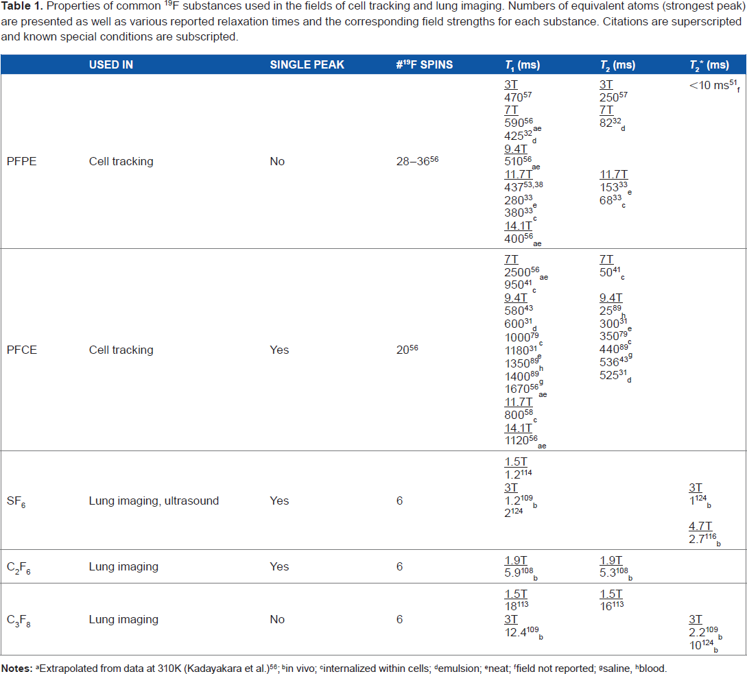
H. Commonly used fluorinated contrast agents that are used to probe the microbiological and macrobiological environments include perfluorocarbons (PFCs) such as perfluoropolyether (PFPE), perfluoro-15-crown-5-ether (PFCE), sulfur hexafluoride (SF6), perfluoropropane (C3F8), and hexafluoroethane (C2F6). Their usage and important physical properties are conveniently summarized in Table 1. Fluorinated contrast agents are used to probe the microbiological environment as cell-labelling agents
4
and theranostic drugs and therapies. Therapeutic delivery of labeled cells using PFC emulsions (eg, dendritic cells [DCs]
4
) or drugs (eg, 5-fluorouracil
5
) containing
19
F can be quantified and tracked noninvasively and longitudinally using MRI. Agents such as fluorinated gases are also useful for probing macrobiological environments such as the airspaces of lungs.
6
Inert fluorinated gases containing multiple chemically equivalent
19
F atoms per molecule are used in the field of MRI of the lung as inhaled contrast agents to probe lung structure and function noninvasively. This article discusses the role of
19
F in the fields of cell tracking and lung imaging. Many new contributions have been reported in the past few years in these fields and their importance will be discussed. Though drug development is a large contributor to the
19
F body of literature, its scope is vast, and we direct interested readers to the thorough review on the role of
19
F in imaging and drug development by Bartusik and Aebisher.
7
Publication frequency by year for “
19
F and MRI” on PubMed. Additional search parameters are included in the legend separating out the fields of cell tracking, lung imaging, and drug development. The rate of publications for
19
F-based cell tracking is observed to increase over the past decade since the initial demonstration of the concept in 2005.
4
Properties of common
19
F substances used in the fields of cell tracking and lung imaging. Numbers of equivalent atoms (strongest peak) are presented as well as various reported relaxation times and the corresponding field strengths for each substance. Citations are superscripted and known special conditions are subscripted.
Extrapolated from data at 310K (Kadayakara et al.)
56
in vivo internalized within cells emulsion neat field not reported saline blood.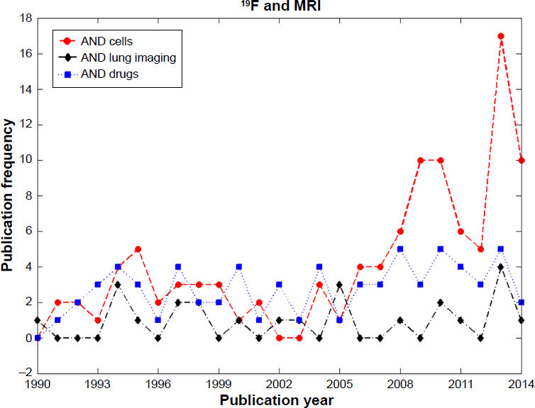
Cell Tracking Using Imaging Techniques
Cell tracking is an important tool for the field of cellular therapy. The goals of cellular therapy are to stimulate the immune system to elicit an immune response, eg, in cancer patients, and regenerate damaged tissues using stem cells. The requirement and need to track cells noninvasively is of great importance for the development of cellular therapies and vaccines for eventual clinical translation and acceptance. Therefore, cell tracking is a necessary tool in order to help answer the questions: How many cells were actually transplanted? How long do transplanted cells survive? Do transplanted cells migrate and reach their therapeutic targets? Though theory can help predict the status of cells and routes of migration, in practice, the outcomes are often very different and patient specific, which is why methods of noninvasive imaging are needed. Previously developed cell-tracking techniques employed the use of fluorescent labels, radio labels, iron labels, and combinations of these for dual-mode identification (eg, iron with fluorescent tags), but all of these techniques have restrictions on clinical feasibility, which will be described in the following sections. Fluorinated contrast agents have emerged as a recent addition to these techniques in the form of PFC emulsions and other fluorinated compounds.
It is important for any cell-labelling strategy that the substances used as labelling agents do not alter the properties of cells, do not cause cytotoxic effects to the cells, and do not change their phenotype or behavior. Using fluorescence-activated cell sorting (FACS), consistency of cell phenotype can be verified aiding in a novel label development. Simple assays using trypan blue can be used to compare cell viability with and without label to reveal any negative cytotoxic effects of the label on therapeutic cells. Practical issues arising from cell label retention are cell division, cell death, and the potential for bystander cells to adopt labels. Cell division leads to a dilution of contrast agent content per cell, reducing their individual detectability for highly localized labeled cells. Upon cell death, the label is free to leak out from the cells and become dispersed. Bystander cells may phagocytose the recently released label from dead cells and contribute to false-positive results. Important clinical translation considerations include the safety profile of the labelling agents, whether there are any toxic effects, and how the labels are metabolized and cleared from the body (biological half-life) once they leave the cells they initially resided in.
Nonfluorinated Cell-Tracking Methods
Previously developed cell-tracking methods include the use of fluorescence microscopy, positron emission tomography (PET) using radioactive agents to label cells, and MRI using heavy metal contrast agents (eg, superparamagnetic iron oxide [SPIO], Gd). Fluorescence microscopy using fluorescently tagged cells has the ability to reveal tagged cells in organs with enough resolution and signal-to-noise ratio (SNR) to resolve even single cells. However, the ability to observe cells in vivo using these methods becomes difficult as tissue depth increases, limiting in vivo studies to small animals. A thorough review of fluorescence molecular imaging can be found here. 8
The use of radio labels in single-photon emission computed tomography (SPECT) and PET provides high sensitivity and has been used for cell-tracking applications.9–11 There are some significant drawbacks to these techniques, which include the following: their radioactive nature, which imparts ionizing radiation to cells and tissues, resulting in potential cytotoxicity to the cells they label; relatively short nuclear half-lives, limiting the effectiveness for longitudinal studies; and limited spatial resolution. However, the latter may be overcome by recent developments in modern combined MR/ PET or PET/computed tomography (CT) systems to provide improved localization of cells due to high-resolution anatomical imaging from their MR and CT counterparts.
Cell tracking using iron labels was first utilized by Bulte et al and Yeh et al in the early 1990s by internalizing dextran-coated magnetite particles and SPIO particles (Aquamag100 and BMS 180549) within cells and detecting them using MRI.12–14 Iron-labeled cells are usually identified by the negative contrast observed in MR images due to their enhancement of
Fluorinated Contrast Agents for Cell Tracking Using MRI
The use of PFCs for cell tracking using MRI was first demonstrated in 2005 by Ahrens et al describing a complete imaging platform for tracking immunotherapeutic cells.
4
PFC-labeled cells provide direct detection of cells in the form of positive-contrast
Labelling of cells using PFCs can be performed using ex vivo methods (cell culture flasks) or in situ as an injection (usually intravenous). For the ex vivo case, cells are incubated at a specific concentration of cells (typically 2 × 10 5 cells/mL) and 19 F-containing label within the cell media in order to promote cellular uptake. Uptake is not instantaneous, and there is an optimum label dose and incubation time depending on the agent used and due to natural cell division during the labelling period. Potential cytotoxic effects can occur due to extended exposure to high concentrations of label within the media. This is further complicated by the fact that these optima are different for each cell type of interest. These parameters are important to map out to maximize the cellular uptake of label (which translates to improved SNR within post transplant images) as well as to ensure that the labels do not affect the cells’ behavior (eg, migration, differentiation, and surface marker expression). One method to increase the available SNR within the cells is to increase the number of 19 F spins by means of using larger label particle sizes. However, it has been shown that there is an optimum size where smaller sized particles (<560 nm) do not disrupt cell behavior. 25 Labelling cells ex vivo is a very powerful technique owing to the ability to label specific cells in controlled conditions with the potential to quantify absolute counts of cells.
Cell labelling in situ is more straightforward but less target specific and lacks the potential to quantify cell numbers absolutely. To label cells in situ, an injectable PFC emulsion is formulated and administered intravenously. While within the vascular system, circulating cells (monocytes, macrophages, etc.) phagocytose the label and become PFC-labeled cells. Cells labeled this way usually consist of a distribution of cell types possessing variable label concentration. This technique is often used to label the influx of inflammatory macrophages responding to a recent insult.26–31
Noncommercial and Commercial Agents for Cell Tracking
There are a variety of commercial and noncommercial fluorinated labelling agents currently available for use for cell labelling. These include linear PFCs such as the PFPE Cell Sense (CS-1000, CS-1000 ATM and fluorescently tagged CS-ATM DM Red, Green, NIR),32–38 V-Sense (VS-1000H),27,39,40 and cyclic PFCs such as PFCE-based agents,29,41–44 including VS-580H, which is a commercial PFCE,45–47 as well as other PFC formulations such as perfluorooctyl bromide (PFOB), 31 perfluorodecalin (PFD), 31 trans-bis-perfluorobutyl ethylene (F-44E), 31 and superfluorinated compounds (eg, PERFECTA 48 and 19 F I T. 49 ) These latter formulations aim to provide more SNR per cell (by fitting even more 19 F atoms per molecule within the labels while maintaining small particle sizes) and adding additional functionality such as the ability to cleave the molecule apart for easy clearance by enzymatic action.48,50 Additional information on the hydrodynamic diameter and specific target of many PFC agents has been well summarized in previous reviews. 51
At the time of writing this article, Cell Sense and V-Sense are the two commercially available PFCs used for cell tracking in MRI, which are manufactured by Celsense Inc. They have developed several formulations of ex vivo and in situ cell labels, which were originally filed for patent on May 2, 2008 (no. PCT/US2009/002706). These include fluorinated emulsions (CS-1000, CS-580, VS-1000H, VS-580H) as well as other formulations that incorporate fluorochromes (CS and VS–-ATM DM Red, Green, and NIR) for detection with MRI and fluorescence microscopy (ie, dual-labeled cells). Formulations using the numerical suffix “1000” are composed using PFPE, whereas those using “580” are composed from PFCE. To our knowledge, this company manufactures the only Food and Drug Administration-approved PFC label suitable for clinical cell tracking (ie, CS-1000) providing groundwork and a desirable research route for translational cell tracking and cellular therapy clinical trials. The safety of some PFCs in biomedical applications has been investigated previously. The PFC PFOB has been characterized and used as a blood substitute in large quantities.24,52 The amounts of PFC used as in situ and ex vivo agents for cell tracking are quite small in comparison with the quantities used as blood substitutes. This reduces the concern of residual label and clearance over time. Clearance of PFC from the body is primarily achieved via the reticuloendothelial system and the lungs. 52
Cell Quantification from Imaging
The ability to quantify the MRI signal obtained from
19
F images of PFC-labeled cells is paramount to the development of future cellular therapies leading to conclusions about cell fate after administration. Ex vivo techniques allow for the quantification and potential to measure absolute cell counting from MR images. The processes involved are depicted in Figure 2 and are as follows. Cells are first labeled by incubating them with a
19
F-based labelling agent (typically 48 hours). Three important measurements are required for quantification: The fractional PFC content per cell, denoted Fc, the SNR obtained from a uniform reference phantom (typically a dilution of the original cell labelling agent), and the SNR obtained from the regions of interest containing the cells themselves. Obtaining Fc is an extra step using a small fraction of the labeled cells separated from the transplant batch, as shown in process 1. To calculate Fc, this known quantity of cells is characterized using NMR and its spectrum is compared with a reference possessing a known concentration of
19
F within the same NMR tube (trifluoroacetic acid [TFA]). Process 2 involves obtaining conventional
1
H MR images in animal models or human beings after the injection of
19
F-labeled cells to obtain the underlying anatomy at high resolution. Finally, in process 3,
19
F MRI is performed depicting
19
F signal from the reference phantom as well as the injected labeled cells. Obtaining the SNR values from injected cells and the reference phantom within these images, together with Fc, enable the estimation of the number of cells detected within an image. These methods are described in detail by Srinivas et al.
53
Numerous in vitro quantification studies using this method have reported a Pearson correlation coefficient ( This figure depicts the ex vivo labelling process for absolute labeled cell quantification. During the cell-labelling phase, cells are first incubated with a
19
F-based agent for up to 48 hours in order for
19
F spins to become internalized within cells. Labeled cells are then utilized in three different processes. Process 1 demonstrates the performing of NMR on a small sample of these labeled cells to obtain their fractional fluorine content (Fc). Process 2 involves conventional
1
H imaging of animals and human beings post injection to provide the underlying anatomy. Process 3 involves
19
F imaging of animals and human beings to provide signals from both a reference phantom of known concentration and signals from the injected cells. The signals from these images, combined with Fc, allow for quantification of injected cells.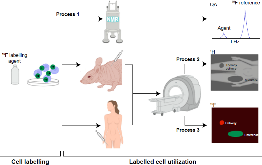
Physical Properties, Hardware, Pulse Sequences, and Reconstruction
In addition to the various labelling agents available, raw numbers of 19 F per molecule, relative chemical shifts, MRI hardware, and pulse sequence development play an important role in increasing the sensitivity and detectability of PFC-labeled cells.
Important physical properties of commonly used PFCs are their spin–lattice (
Research utilizing PFCs for cell tracking requires additional hardware not available on all scanners. Broadband amplifiers capable of nonproton excitation are generally required as are dedicated radiofrequency (RF) coils capable of detecting the 19 F resonance. The separation between 1 H and 19 F frequencies is small compared with that of other nuclei (~4–30 kHz between 1.5 and 11.7 T). For clinical field strengths, existing amplifiers may be sufficient for the detection of 19 F signals without the need for an expensive multinuclear package. RF coils capable of detecting both 1 H and 19 F signals are desirable for both preclinical and clinical imaging. Some examples include single-tuned linear coils and dual-tuned coils that can be manually or electronically switched between scans. 57 A big advantage of dual-tuned coils is that switching between 1 H and 19 F modes can be performed without having to remove the sample, animal, or patient from the bore. 54
Together with these specialized RF coils, MRI pulse sequences are needed. The majority of reported work using PFCs for MRI cell tracking utilized fast spin-echo pulse sequences such as rapid acquisition with relaxation enhancement.4,27–29,32,33,40,42,43,45,46,59,60 Due to the refocusing pulse, this sequence has the advantage of decreasing
Image reconstruction considerations are important as well. Detecting small populations of PFC-labeled cells does not yield exceedingly high SNR values (~5–15), and SNR thresholds (to distinguish real signal from background) are typically quite low (between 2.5 and 5). Postprocessing is required for low SNR data by performing Rician noise corrections53,64 and for removing artifacts caused by chemical shifts.
65
Compressed sensing is an imaging method that uses sophisticated image reconstruction to form images from sparsely filled
Applications of PFC to Cell Tracking
Since 2005, PFC cell tracking has been applied to numerous cell types and disease models, including the tracking of endothelial cells, 55 inflammation (macrophages),26–29,31,42,45,47,59,70–73 monocytes,30,39,74 cancer cells,75–78 human-derived mesenchymal stem cells (hMSCs),36,37,61 stem/progenitor cells, 58 DCs,4,25,34,41,44,79,80 T-cells,38,53,78,81 hematopoietic stem cells, 35 neural stem cells,32,33,43 and natural killer cells. 82
Mesenchymal stem cells (MSCs) are of recent interest in cellular therapy due to their ability to modulate various immune responses 83 and their capability of differentiating into various tissues such as bone, cartilage, and adipose. 84 Not surprisingly, PFC cell tracking has been recently used to investigate mouse-derived MSC (mMSC) and hMSC.36,37,61 The development of hardware and imaging protocols in these animal models is essential to lay the groundwork for future clinical translation.36,37,61 A recent study investigated the fate of MSC implants labeled using CS-1000 in two mouse transplant models using immunocompetent and immunocompromised mice. 61 mMSC (2 × 10 6 ) and hMSC (1.5 × 10 6 ) were delivered to competent and compromised mice, respectively, and were imaged at four time points for up to 17 days. For both mice, day zero quantification agreed well with the expected implanted cell numbers. Over time, 19 F signal decreased, and in the immunocompromised mice, it decreased at a slower rate than those with immunocompetent systems. Results were consistent with the performed immunohistochemistry. Here, imaging with 19 F-MRI provided an advantage over traditional postmortem tissue analysis by providing longitudinal cell information from the same animal. Another point of impact of this work is the fact that this is the first detailed report of the problem of the use of isoflurane as an anesthetic for preclinical cell tracking using PFCs.
Isoflurane and 19 F Cell Tracking
Isoflurane is a fluorinated anesthetic commonly used in experiments involving animal models and has the potential to corrupt signals originating from PFC-labeled cells due to buildup within fat/adipose tissue and potentially from within the lung. Isoflurane has two resonances that are very close to the resonances of PFCs commonly used in cell tracking (PFPE and PFCE) with values of -6.8 and -13.9 ppm relative to TFA (eg, PFPE is at -16 ppm relative to TFA).
85
A table of isoflurane chemical shifts relative to TFA in common solvents can be found here.
86
Relaxation times of these peaks depend on field strength and their local environments.
The problem of isoflurane detection in animal models of PFC cell tracking is not widely discussed in the literature and where it is, mere avoidance of the
19
F signals from isoflurane is mentioned by using alternate anesthetics. Furthermore, image acquisition parameters helpful for pulse-sequence design to avoid exciting isoflurane are seldom given (eg, RF pulse shape and duration). Boehm-Sturm et al described the use of ketamine/xylazine maintained by syringe pump for their animal experiments to avoid the
19
F signals due to isoflurane.
33
van Heeswijk et al also acknowledged isoflurane as a potential problem and presented spectra showing peaks due to isoflurane in close proximity to PFCs, albeit at a signal strength an order of magnitude lower than those of the PFCs.
89
Ribot et al recently utilized pentobarbital rather than isoflurane to avoid isoflurane signal.
37
The first reported use of isoflurane in experiments involving cell-tracking applications was in 2010 in rats
76
and mice.
34
Before that, ketamine/xylazine was the preferred method of anesthesia presumably to avoid detection of isoflurane. Today, isoflurane is used extensively, even in cell-tracking applications, likely attributed to its ease of use in implementation.25,27,30,39,40,46 With these reports and physical attributes in mind, it would seem that isoflurane is still very difficult to detect and may not be worth worrying about. However, working toward clinical translation, animal models will have to move to lower field strengths (≤3 T, reducing spectral separation in Hertz). This may require increases in scan time resulting in an increase in isoflurane deposition in fat due to reduced PFC SNR. More efficient imaging techniques will also increase sensitivity to low-density
19
F sources due to efficient use of SNR/t and shorter TE (eg, TE 1.8 ms
61
), which is simultaneously beneficial for detecting signals from both cells and isoflurane. Under these conditions, attention to isoflurane may be required using solutions such as shaped RF pulses employed to exclude excitation of unwanted peaks
61
or reversion to nonfluorinated anesthetics for animals. Figure 3 demonstrates the improvement (reduction of isoflurane signal) in
19
F images when considering excitation pulse parameters such as shape and pulse duration.
(A) Strong isoflurane signal (red arrow) is detectable following accumulation in the fat pads of mice after excitation with the standard Gaussian filtered sinc pulse (B). This signal is chemically shifted from the fat pad (blue arrow), compared to that of the reference tube above. (C) The mouse was then scanned with a non-filtered sinc pulse (D). Fourier transform of this pulse produces a narrower excitation that did not excite isoflurane
19
F atoms, reducing unwanted background signal from isoflurane. Both images have been windowed to the same level, and brightened to show the noise distribution. (B) The filtered pulse shape in time space is shown, with a width of 0.66 ms. (D) The non-filtered sinc pulse width is much broader with a FWHM of 1.32 ms in time space. image courtesy of Gaudet et al
61
and reproduced with permission.
Clinical Cell Tracking
The first use of PFC-labeled cell tracking in human beings was recently demonstrated by Ahrens et al, who described the results of a phase I clinical trial in the United States using a DC vaccine designed for stage 4 colorectal cancer treatment.
57
Since the strength of treatment is proportional to the number of migrating DC, it is essential that researchers have a noninvasive, quantitative technique to monitor therapy. In addition, since lymph node removal is not practical in the clinical setting, imaging provides the only available information for dose optimization. Five patients were enrolled and assigned into groups of low dose Result of the processes described in Figure 2 as applied to human cell tracking. In vivo MRI in patients following intradermal DC administration into quadriceps. in these patients, ~1 × 10
7
labeled cells were injected. (A) A representative
19
F Magnetic Resonance Spectroscopy (MRS) spectrum of the patient at four hours postinoculation. The DCs appear as a single narrow peak. “Reference” is from an external tube containing TFA placed alongside the patient. (B) axial composite
19
F/
1
H images of the right thigh at 4 hours postinoculation in three patients, a 53-year-old female (left), a 45-year-old female (middle), and a 61-year-old male (right), where DCs are rendered in 
Lung Imaging Using Inert Fluorinated Gas MRI
19 F-based MRI has a role not just in imaging microbiological environments (cell tracking) but also within macrobiological environments, such as the lungs’ airspaces. Lung MRI using inert fluorinated gases as contrast agents is another field enjoying recent success due to technological advancements for 19 F detection. Hyperpolarized (HP) noble gas MRI of the lung is a field that is >20 years old, born out of the desire to study the effect of anesthesia on brain function. The first HP gas lung image was obtained using MRI and HP 129 Xe in excised mouse lungs. 90 Since then, this field has flourished initially due to the success and ease of use of HP 3 He lung imaging. Together, these nuclei have been used to investigate and address various chronic respiratory diseases and acute lung injuries in both clinical studies and animal models, forming the basis of HP noble gas lung imaging. These gases have the ability to provide anatomical and functional information from within the lungs of human beings and rodents such as static ventilation imaging, 91 characterization of apparent diffusion coefficients (A DCs),92–96 and gas exchange, including those from models of emphysema, asthma, fibrosis, radiation-induced lung injury, and inflammation due to fungal spores.
HP gas MRI of the lung requires the use of expensive polarizers to achieve high SNR within the lung. In hyperpolarizing these isotopes, they receive enhancement of their nuclear polarization by a factor of 1 × 10 5 compared to their thermal equilibrium values resulting in their high SNR and excellent image quality. Furthermore, the isotopes used are also expensive and, in some cases, are becoming increasingly scarce (eg, 3 He). 3 He has been used quite extensively over the years; however, within the past decade, interest has shifted toward 129 Xe due to improved polarization techniques for 129 Xe,97–99 ease in obtaining these isotopes, and ability to perform novel functional measurements such as gas exchange at the blood gas barrier,100–102 perfusion measurements, 103 and brain function 104 due to 129 Xe's high solubility in tissues and blood compared with 3 He. 129 Xe has a natural abundance of 26% and small gyromagnetic ratio (~28% of 1 H). Though xenon is a valuable by-product of the liquid air industry and is quite easy to obtain, mixtures enriching the 129 Xe isotope can greatly increase SNR but at a much higher monetary cost.
Fluorinated gases require no such polarizer, are less costly to purchase, and the signal is derived from its thermal equilibrium polarization, just like conventional 1 H MRI. In addition, these gases comprise molecules possessing multiple NMR equivalent fluorine atoms collectively enhancing their detectability. The most common fluorinated gases used to date for lung imaging (especially in human beings) are SF6 and C3F8. C2F6 has also been studied extensively in animal models and use of tetrafluoromethane (CF4) has also been reported. These are not rare gases unlike 3 He and 129 Xe. Furthermore, use of these gases for MRI enables safe and repeatable imaging that is noninvasive, nonionizing and there is no 19 F background.
The first 19 F gas lung images were obtained in 1982 by Heidelberger and Lauterbur using CF4 in excised rabbit lungs, 105 preceding the first HP gas lung images by 12 years. Shortly after, images were obtained in excised healthy canine lungs.106,107 After a long gap in 19 F MRI lung reports, Kuethe et al demonstrated continuous breathing of C2F6 in rats, 108 and it was not until 10 years later that the first human lung images were reported by Wolf et al 6 using a mixture of SF6 and oxygen. Though these gases currently produce images that are lower in quality compared with HP gas images, they come with many benefits, as described below. These gases are important because they provide similar diagnostic information and biomarkers as HP gases (eg, static ventilation images,6,108–111 ADC,112–117 and surface-to-volume ratio 114 ) and complement them as alternative methods to probe healthy and diseased lungs.
Inert fluorinated gases are not the only methods available to perform MRI of the lung without the use of HP gases. For completeness, oxygen-enhanced methods, 118 Fourier decomposition, 119 and 1 H UTE 120 exist. Recent comprehensive reviews of lung imaging using MRI have been presented.121–123
Physical Properties, Hardware, Pulse Sequences, and Reconstruction
Of the commonly used fluorinated gases (ie, SF6 and C3F8), there are several key physical properties that impact their effectiveness in the MRI of the lung. These include their relaxation times, spectral features, number of equivalent 19 F atoms, and diffusion properties. Table 1 summarizes the key properties for these gases and C2F6.
In general, the longitudinal relaxation times for these gases are short (1–30 ms). Bulk transverse relaxation times are even shorter with values less than a few milliseconds. SF6 and C3F8 are recently reported gases used for human lung MRI. Just like their cell-tracking counterparts (PFCE and PFPE), SF6 and C3F8 possess one resonance and two resonances, respectively. SF6's single peak arises from six equivalent
19
F atoms with reported
Unlike PFCs used for cell tracking, the diffusion coefficient of these 19 F gases plays an important role for probing lung microstructure and performing functional lung imaging. The theory and use of 19 F gases for diffusion-weighted MRI is extensively described in these reports.112,113,115–117
Fluorinated gases have a good record of safety due to their inertness, supported by their use in recent human imaging studies as inhaled contrast agents and lack of reported adverse events in the literature. Furthermore, these gases can be directly mixed with physiological concentrations of oxygen (21%) without large penalties in SNR, unlike HP gas agents. 113 The ability to create normoxic mixtures eliminates the risks involved with long-duration breath-holds of anoxic gas volumes and enables repeated breathing and even washout maneuvers in order to reach a steady state of 19 F gas within the lung as well as perform dynamic lung imaging.
The hardware challenges for 19 F lung imaging are similar to those for 19 F cell tracking. Amplifiers capable of excitation for a range of 19 F resonances (~150 ppm for 19 F gases 114 ) are required as are dedicated RF coils capable of detecting the 19 F resonance. Lung imaging is commonly performed at clinical field strengths between 1.5 and 3 T. Imaging may prove difficult at higher field strengths due to the high magnetic susceptibility of lung tissue at the air–tissue interfaces. In fact, the theoretically predicted optimum field strength for HP gas human lung MRI is between 0.1 and 0.6 T. 125 Specific absorption rate (SAR) depends on the field strength and may impose additional safety restrictions at field strengths >3 T. RF coils capable of detecting both 1 H and 19 F signals are desirable for both preclinical and clinical imaging, though designs exist that allow the use of the scanners’ native body coil for 1 H by actively detuning the 19 F coil without removal of the coil or patient.109,110,126 Furthermore, the spatial extent of 19 F signals is quite larger in comparison with cell-tracking applications (eg, 5 L lung volume vs. 1 mL injection sites) requiring different RF coil solutions such as large volume coils of birdcage design or flexible surface coils. Unlike cell tracking, which for some applications requires minimal penetration depth (~1–2 cm), lung imaging requires uniform excitation of the entire lung, requiring high transmit uniformity and power. Birdcage designs have been employed for 3 He,91,127 129 Xe, 128 and 23 Na, 129 but to our knowledge, no 19 F coils of this design have been reported. The use of a coil of this type for 19 F lung imaging would provide high B1 uniformity and require less effort in setup from patient to patient. Other reported coils used for 19 F lung imaging employ flexible volume coils with a detuning circuit to allow for 1 H body coil imaging.109,130 This design allows for easy imaging between 1 H and 19 F frequencies and also provides patient comfort. Due to the flexibility of the coil, however, B1 mapping is required on a case basis in order to correct the images for inhomogeneous B between patients.
Due to the short relaxation times of these gases, MR pulse sequences having short repetition time (TR) and TE are desirable. The shorter TR reduces the overall scan time providing a time-saving that can then be used to enhance SNR by rapid signal averaging (subject to specific absorption rate limitations). Several sequences used for imaging
19
F gases have been described in the literature, including 3D gradient recalled echo (GRE),110,130,131 2D/3D FLASH,6,132 UTE,
109
COMSPIRA,94,116,117 and X-centric, a type of GRE.
124
UTE and 3D GRE methods have been compared demonstrating improvements in SNR for UTE at the cost of edge resolution.
131
X-centric has also been compared to UTE and GRE methods in a resolution phantom demonstrating both improvements in SNR and sustained resolution compared with UTE and GRE at the cost of a second scan (because only 50.5% of
Isoflurane and 19 F Lung Imaging
Though isoflurane is in common use as an anesthetic for animal models used in cell tracking, there is no description of isoflurane posing a problem for 19 F lung imaging. This is likely due to the fact that the described preclinical research in rats and pigs all used injectable anesthetics such as sodium pentobarbital, ketamine, xylazine, and thiopental rather than inhaled anesthetics such as isoflurane and halothane to deliberately avoid 19 F signals from their anesthetics.
Static Lung Imaging
Static ventilation lung imaging is the easiest and most common method of lung imaging to perform. In general, a volume of inert fluorinated gas is introduced to the lungs using a ventilator (for animals) or inhaled from a plastic bag (for human beings) as depicted in Figure 5. In either case, once the dose is delivered, the animal/human being engages in a breath-hold and MRI is performed during a modest breath-hold length of up to 15 seconds. During the breath-hold, motion artifacts or blurring due to breathing are of no concern due to the temporarily suspended respiratory motion. The gas is then exhaled and the experiment can be repeated. In some cases, the volume of the lung is washed out using prebreaths of
19
F gas before the imaging breath-hold is initiated in order to provide a uniform distribution of inert fluorinated gas to all possible air spaces. There may be inherent differences in lung volume between
19
F images and
1
H due to the fact that separate breath-holds are needed for each. These differences are controlled to the best of the researcher's ability (eg, functional residual capacity (FRC) + 1 L of
19
F gas or air). The first
19
F gas lung images obtained by Heidelberger and Lauterbur in 1982
105
as well as those demonstrated by Rinck106,107 and Keuthe,
108
are the first examples of static lung imaging.
Medical-grade inert fluorinated gases are provided by compressed gas companies and are ready to use straight from the cylinders unlike HP
3
He and
129
Xe. Animals and human beings are administered these gases either using a specialized animal ventilator or from bags containing large volumes of gas (1–5 L), which patients inhale from. During a breath-hold, imaging of the inert fluorinated gases is performed, which reveals information of the ventilated airspaces of the lungs.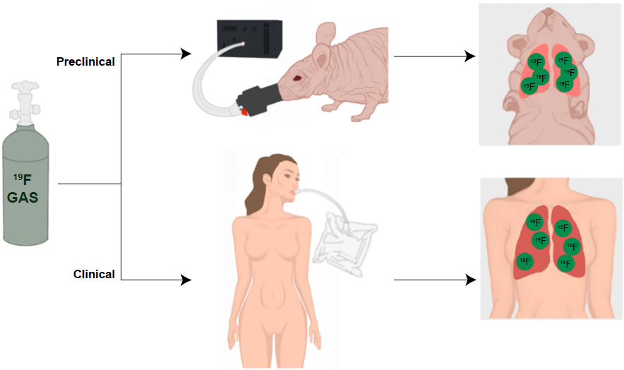
Dynamic Lung Imaging
Dynamic lung imaging using fluorinated gases was first described by Schreiber et al using SF6 in pig lungs to measure wash-in and washout kinetics using dynamic gradient echo imaging. 133 Kuethe et al measured the volume of rat lungs during the respiratory cycle of mechanically ventilated rats using SF6 in a disease model of elastase-induced emphysema. 134 By calibrating NMR signal strength to known volumes of SF6 within the laboratory, fast spectroscopic techniques can be used to measure in vivo lung volume with high temporal resolution (20 measurements per second). Wolf et al investigated MRI of dynamic wash-in of fluorinated gas using C2F6 in pig lungs to optimize image acquisition for very short acquisition times with respectable image quality in as short as two seconds. 132 They also investigated C3F8 in a lung washout study and, for the first time, demonstrated and quantified washout dynamics under high-frequency oscillatory ventilation. Scholz et al compared 19 F gas-derived measurements of lung function and did a direct comparison with nonimaging respiratory gas analysis measuring alveolar gas fraction in pigs. Ouriadov et al investigated and compared ventilation mapping using SF6 and PFP after establishing improved resolution and SNR using the X-centric pulse sequence compared with UTE and fast gradient recalled echo (FGRE) within a resolution phantom. X-centric can achieve relatively short TE (~0.5 ms) while maintaining the benefits of Cartesian style acquisition. 124
Functional Lung Imaging (Diffusion, paO2, Fractional Ventilation, and Gradients)
Expanding on the possible detectable biomarkers using fluorinated gases for lung MRI, ADCs of gases,114–117,135 oxygen partial pressure (paO2), ventilation perfusion (
Clinical Lung Imaging
The first human lung images obtained using
19
F gas were reported by Wolf et al
6
using a mixture of SF6 and oxygen. Recent static ventilation imaging in human beings has been reported using enhanced techniques to overcome the low sensitivity of
19
F gas lung imaging. The use of a large pre-washout of the lungs by inhaling several breaths of fluorinated gas mixed with 21% oxygen from bags
130
or delivery system (Physiorack)
126
greatly increases the number of
19
F spins within the lungs prior to imaging. Couch et al utilized a large 5 L bag of gas (C3F8 or SF6 mixed with 21% of oxygen) to washout lungs prior to inhalation of 1 L of gas from a second bag to ensure imaging was carried out at FRC + 1 L.
109
They employed an UTE imaging sequence (3D UTE) and compared the results with the previously reported FGRE images reporting a factor of 2 enhancement in SNR. Representative UTE images are shown in Figure 6.
Coronal pulmonary
19
F 3D UTE MR images obtained from a healthy volunteer during a 15 second breath-hold after continuous breathing from a 5 L mixture of 79% PFP and 21% oxygen followed by a 1 L inhalation of the same mixture in order to image lungs at FRC + 1 L with an equilibrium distribution of the inert gas mixture. Image courtesy of Couch et al
109
and reproduced with permission.
Halaweish et al performed the first 19 F enhanced dynamic imaging studies on healthy volunteers and volunteers with emphysema and chronic obstructive pulmonary disease, demonstrating differences in ventilation heterogeneity and gas trapping in those with chronic obstructive pulmonary disease. 110 Due to the short relaxation times of these fluorinated gases, sequences employing shorter TEs can greatly impact SNR in a beneficial way. Finally, measurement of surface-to-volume ratios 114 and gravitational gradients 114 have also been demonstrated in human lungs using PFP; however, a full study comparing healthy lungs with diseased lungs remains to be reported.
Conclusions
The use of fluorinated contrast agents for MRI of cell tracking, lung imaging, and drug development represents a large body of scientific research. Various problems and solutions, owing to the range of field strengths (1.5–11.7 T) that this research spans across, the relaxation properties of different fluorinated contrast agents, and practical issues have been described. UTE strategies, isoflurane usage, and detection of 19 F agents at clinical field strengths are where cell tracking and lung imaging may mutually benefit each other. Cell tracking may benefit from the development of UTE strategies for lung imaging, which may improve detection of 19 F-labeled cells in the presence of iron for multicell population labelling (eg, Hitchens et al 62 ). Preclinical 19 F lung imaging does not utilize isoflurane at all. Alternate anesthesia strategies would eliminate the problems that isoflurane may cause for cell tracking. With clinical translation in mind, cell tracking may greatly benefit from technological advances in lung imaging since the majority of research in that field is performed at 1.5 and 3 T. Novel RF coils and arrays have been developed as well as pulse sequences designed to detect low concentrations of 19 F. Fluorinated contrast agents are indeed versatile contrast agents well suited for probing the micro- to macrobiological environments, and future work will only enhance their utility in these important fields.
Author Contributions
Wrote the first draft of the manuscript: MSF. Contributed to the writing of the manuscript: MSF, JMG, PJF. Agree with manuscript results and conclusions: MSF, JMG, PJF. Jointly developed the structure and arguments for the paper: MSF, JMG, PJF. Made critical revisions and approved final version: MSF, JMG, PJF. All authors reviewed and approved of the final manuscript.
Footnotes
Acknowledgment
The authors thank Chelsey Foster for authoring figures 2 and ![]() .
.