
Abstract
Sulfur mustard (SM), is an alkylating agent and has been emerged as a chemical weapon in various battlefields. More recently, SM was employed in the Iraq conflict against Iranian military forces and civilians. Nowadays there are more than 40,000 people suffering from pulmonary lesions special chronic obstructive pulmonary disease (COPD) due to mustard gas in Iran. SM causes the endogenous production of reactive oxygen species (ROS).
Heme oxygenases (HOs) are the rate-limiting enzyme for heme metabolism. Numerous studies have confirmed that HOs are concerned in diverse biological processes such as anti-oxidation.
The present study was undertaken to consider the regulation of HO-1 and HO-2 n the human airway wall, and to suggest a probable role that HOs may play in cellular defense against oxidative stress due to SM.
In this research ten unexposed SM individuals and twenty SM exposed patients were included. Evaluation of HO-1& HO-2 expressions in unexposed and SM exposed patients samples was performed by semiquantitative RT-PCR, real-time RT-PCR and Immunohistochemistry analysis.
While unexposed SM samples expressed same levels of HOs, expression level of HO-1was upregulated about 3.58 ± 1.93 folds in SM exposed patients in comparison with unexposed ones, we could not find any difference in expression of HO-2 n two groups. In contrast, Immunohistochemistry results showed negative HO-1 protein expression in SM injured patients.
Our results revealed that HO1 may plays an important role in cellular protection against oxidative stress due to mustard gas toxicity in airway wall of SM exposed patients at mRNA level, but translational modifications might cause decrease in the amount of HO1 protein.
Background
Sulfur Mustard (SM) or bis-2-(chloroethyl) sulfide, has been emerged as an important chemical weapon and is classified as a destructive warfare agent. 1 It has been used in several battlefields around the globe and was employed during World War I for the first time.1–3 Since then, within the Iran-Iraq war of the 1980s, there was extensive usage of SM by Iraqi ex-regime against Iranian civilians and military forces. 4
This agent causes acute and late complications which have their own characteristic features. 20 years after encountering SM, side effects of initial exposure to SM, is able to produce severe damage among exposed individuals, mainly in skin, eyes, lungs and respiratory tracks.5,6
To date there are more than 40,000 people with different degrees of involvement undergoing late respiratory complications due to SM exposure. 7 There is no general agreement about the pulmonary pathophysiology of the patient population who were exposed to SM, but chronic bronchitis, chronic obstructive pulmonary disease (COPD), asthma and bronchiolitis obliterans (BO) are the most abundant long-term respiratory diseases as the underlying causes.4,6,8
Despite much research, the precise mechanism by which SM causes late respiratory diseases is still poorly understood. SM as an alkylating agent able to interact with nucleophilic functional groups such as amino, carboxylic and hydroxyl in DNA which refers to the mutagenic effect of SM and stable alteration in genome.3,6,9 Beside the alkylation of DNA, another theory for the late effect of SM is neutrophil and/or lymphocyte dominant disorder secret proteases and themselves manufacture reactive oxygen species (ROS). 10
These hazardous species are recognized to cause oxidative injury in a number of molecules in cells. The probable harmful effects of these species are controlled by a cellular antioxidant protection which comprise enzymes 9 such as hemeoxygenases (HOs).
HO is a microsomal enzyme accelerating the first rate-limiting stage in the breakdown of heme. It cleaves the a-meso carbon of b-type heme to produce equivalent amounts of biliverdin IXa (BV), carbon monoxide (CO), and ferrous iron.11–14 Two main mammalian HO isoforms have been recognized: HO-1 is a 32-kDa protein and an inducible isoform 15 via various stimuli, remarkably expressed in the liver and spleen. HO-2 is a 36-kDa and non-inducible isoform, and exists in the brain, endothelial cells or testes.11,16,17
Activation of HO, especially HO-1 can also be stimulated by multiple nonheme products 16 such as glutathione depletors, 12 UV irradiation, endotoxin, heavy metals, heat shock, nitric oxide (NO), inflammatory cytokines and ROS.11,13,14 The possible explanation for activity of HO in these cases is that it may function as a vital cytoprotective molecule. 14 HO-1 is induced in the lung during stressful situations to counter with oxidative injury and maintain homeostasis in lungs. 18
Although HO causes reduction of oxidative stress and attenuation of inflammatory reactions due to heme removal, which is a powerful prooxidant factor 11 anti-oxidant activity of HO returns to its byproducts. Indeed the liberated CO induces soluble guanylyl cyclase (sGC), in this way reduces leukocyte adhesion, stops platelet aggregation 19 and inhibits muscle cell proliferation. 20 Biliverdin is promptly converted to bilirubin via Biliverdin reductase 21 which is an effective antioxidant, because bilirubin can scavenge ROS and decrease its production. 14 Ferrous iron released by HO, induces the synthesis of ferritin, the iron storage protein, and cytosolic iron efflux, consequently protects cells from oxidative stress.14,18
By considering the production of ROS in lung injury induced by SM, this research will highlight the roles of HO-1 and HO-2 in pathophysiological cytoprotection of the airway wall at mRNA and protein levels in bronchial biopsies of patients who were exposed to SM in comparrison to unexposed ones. In addition, this finding will be useful to demonstrate a new method of treatment of SM exposed patients with antioxidant medicine.
Methods
Sampling
In this study 20 SM exposed patients and 10 healthy participants were comprised. The exposed patients were individuals who had a documented encounter with SM within the Iran–Iraq war. Tissue was donated by volunteers under conditions of informed consent and this survey was conducted in accordance with a protocol approved by Baqiyatallah Medical Sciences University (BMSU) ethics committee.
Data such as age, gender were obtained, as is shown in Table 1. The average age of exposed individuals was about 43.2 ± 6.4, and the mean age of non-exposed volunteers was approximately 41.3 ± 2.5. Cases with positive history of cigarette smoking, elderly patients, addicts, pre-exposure of pulmonary disease such as asthma, lung cancer or respiratory infections and history of countering to toxic agents were excluded from our study.
Subject.

In respect to obtaining biopsies from the bronchi wall, the flexible fiberoptic bronchoscopy (FFB) (Olympus BF1T, Tokyo, Japan) was performed. Patients were topically anaesthetized by applying 2% Lidocaine. Throughout the bronchoscopy, supplementary oxygen was given. To obtain endobronchial biopsy samples, bronchoscope forceps (Olympus, Tokyo, Japan) were passed through the route of fiberoptic bronchoscopy to reach the segmental and subsegmental carinae. Biopsy specimens obtained were put in Tripure Isolation Reagent (Roche Applied Science, Germany) and formaline as well. Samples in Tripure Isolation Reagent were stored at –80 °C until RNA extraction and samples in formaline were placed in the fridge in order to undertake immunohistochemistry studies.
RNA extraction
Total RNA was extracted in conformity with the manufacturer's recommendations using Tripure Isolation Reagent (Roche Applied Science, Germany). For the first step, bronchi biopsies were homogenated in Tripure Isolation Reagent by the mean of Ultra Sonic Homogenator (Hielscher, Germany). The aqueous phase containing RNA was separated and to avoid contamination with proteins, the lowest fraction of aqueous phase was not incorporated in the total RNA sample. Following the previous stage, isolated RNA was eluted in RNase-free water and the quantity and integrity of RNA were measured by NanoDrop spectroscopy (ND-1000, Wilmington, DE).
Semiquantitative RT-PCR
500 ng of total RNA was reverse-transcribed to create single-strand complementary DNA by SuperScript III reverse transcriptase (Invitrogen, Carlsbad, CA) according to the manufacturer's protocol followed by DNaseI (Invitrogen, Carlsbad, CA) treatment and heat inactivation, to remove any residual chromosomal DNA. 1 µl of the resulting cDNA were validated with PCR in a volume of 25 µl, containing 2.5 µl 10X buffer (Takara, Japan), 5 PM deoxynucleoside triphosphate (Takara, Japan), 0.3 µl rTaq polymerase Enzyme (Takara, Japan) and 10 PM Primer Mix. The interested designed primers are listed below: HO-1 forward, 5'-ATGACACCAAGGACCAGAGC-3', reverse, 5'-GTGTAAGGACCCATCGGAGA-3', HO-2 forward, 5'GGAAACCTCAGAGGGGGTAG-3', reverse, 5'-GTGGCCAGCTTAAACAGCTC-3' and β-actin as a housekeeping gene, forward, 5'-TTCTA CAAT-GAGCTGCGTGTGG-3'; reverse, 5'-GTGTT GAAG GTCTCAAACATGAT-3' was purchased from Bioneer (South Korea). PCR was carried out in the same situation, heat held at 94 °C for 5 min, denaturation at 94 °C for 30 sec, annealing at 57 °C for 30 sec, extension at 72 °C for 1 min (30 cycle) and terminal extension at 72 °C for 5 min. PCR products were separated on a 2% agarose gel electrophoresis and the quantity of bands was visually detectable under UV light after staining with ethidiumbromide.
Quantitative real-time PCR
For real-time quantitative PCR, 500 ng of RT product was used in whole volume of 15 µl consisting 7.5 µl of SYBR Green Premix 2X (Takara, Shiga, Japan) and 10 PM of mix primer by Rotor-Gene RG 3000 (Corbett Research, Sydney, Australia). Thermocycling conditions were; heat hold at 94 °C for 1 min followed by 40 cycles of denaturation at 94 °C for 20 sec, annealing at 57 °C for 30 sec, extention at 72° C for 30 sec. The quantity of each gene expression depends on the cycle at threshold (Ct) in which the fluorescence density in PCR microtube rises above background and was normalized by β-actin as our endogenous reference gene.
Immunohistochemistry
For immunohistochemistry 12 SM exposed patients and 8 control samples were enrolled. We have already described the details of immunohistochemistry technique elsewhere. 22 In summary, airway wall biopsy tissues were placed in 4% buffered paraformaldehyde for fixation for one day. The Sections were incubated overnight with a phosphate buffer comprised 30% sucrose. Tissues, 20 µm in thickness, were cut on a cryostat and incubated with HO-1 antibody at proper density for 12 h at 4 °C. The antibody employed in this study was a rat monoclonal antibody raised against HO of human source (Santa Cruz Biothechnology, Inc, USA) at a dilution 1:200. After incubation with the primary antibody, the Sections were washed with PBS and incubated with biotinylated anti-rat secondary antibody (Santa Cruz Biothechnology, Inc, USA). For immunostaining the tissues were afterward detectable using ABC complex (avidin-biotinylated peroxidase complex) system (Vector Laboratory, Burlingame, CA, USA) with DAB as a substrate.
Statistical analysis
Data were revealed as mean ± SD of fold-changes of HOs gene expression in three independent experiments. SPSS software version 15.0 (SPSS, IL) was employed for statistical analyses. For evaluation of differences in mentioned genes expression between SM-injured group and unexposed group student's t-test was used and P < 0.05 was considered as significant.
Results
Clinical features
In this study a total of 20 subjects who had experienced exposure to SM and 10 unexposed participants were recruited as controls (Table 1).
Expression of the HO isoforms in bronchial wall
It has been shown that two main HO isoforms, HO-1 and HO-2 are involved in degradation of heme. Only HO-1 is an inducible form and is activated via external stimuli11,16,17 Fig. 1 shows the mRNA expression levels of HO-1 and HO-2 in SM exposed and unexposed ones obtained from Semiquantitative RT-PCR. Significant HO-1 expression of SM exposed patients compared to unexposed ones was observed. HO-2 was also demonstrable in all specimens, but levels of HO-2 expression did not show any notable differences between patients exposed to SM and unexposed group.
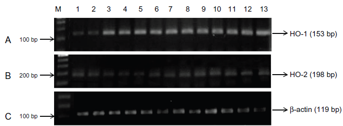
Expression levels of HO-1 and HO-2 in bronchial wall of patients with long term exposure to sulfur mustard. Total RNAs were purified and then analyzed by Semiquantitative RT-PCR. This panel shows gel bands in order to PCR amplification products of HO-1 (153 bp), HO-2 (198 bp) and β-actin (119 bp) transcripts. A) A marked increase in HO-1 expression levels of SM exposed patients (Lanes 3–13) was recognized in compare to expression level of unexposed ones (Lanes 1 and 2). B) The density band of HO-2 in SM exposed patients did not show any highlight deference with mRNA of unexposed group. C) Beta-actin was used as internal control. Lane M shows DNA ladder (100 bp).
To determine the fold changes in expression quantity of target genes, HO-1 and HO-2 mRNA levels were measured by Real-time RT-PCR. Test samples of all exposed to SM elicited 3.58 ± 1.93 fold increase in HO-1 mRNA expression in compare to unexposed group (P = 0.01). But we could not find increased expression of HO-2 mRNA in comparison to unexposed group (P = 0.413) (Table 2).
Increases in HOs expression in SM-exposed patients in comparison with control group.
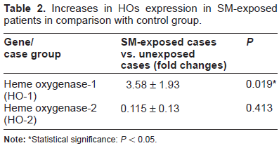
Statistical significance: P < 0.05.
Localization of HO-1 by immunohistochemistry
To consider whether SM causes the expression of HO-1 in protein level, immunohistochemistry was carried out. A basal expression of HO-1 was visible in the bronchial epithelial cells of all members of the unexposed group. The thickness of bronchial epithelium increased in all SM- exposed patients and this may be one of the important reasons for the obstructive condition. HO-1 immunopositivity strongly expressed in epithelial cells of the unexposed group especially in luminal border of epithelium and nucleuses as well (Fig. 2A). In contrast in SM- exposed patients immunoreactiovity for HO-1 in the same cells were negative (Fig. 2B).

Immunohistochemical localization of HO1 in the bronchial epithelium of human airway. A) a section from nonexposed human air way wall that immunostained for HO1. HO1 intensely expressed in the luminal border (Lb) and basal cells (Bc). B) No immunoreactions is seen in the human airway wall exposed to SM. Note to the thickness of SM exposed bronchial epithelium which significantly increased in compare to nonexposed one.
Discussion
In this study we demonstrated that bronchial biopsy of patients with history of exposure to SM was active in upregulating expression of HO-1 at mRNA levels in comparison to unexposed ones, wheras we could not identify any considerable differences in HO-2 transcripts between SM exposed groups and unexposed groups. The main reason for this result is that despite of HO-1, HO-2 is not an inducible isoform and constitutively expressed for the most part of the body.11,16,17
Our findings are approximately consistent with previous studies in that up-regulation of HO-1 mRNA expression has been revealed under oxidative stress situation. As Maeshima showed, HO-1 activity could also be stimulated by a diversity of nonheme products such as heavy metals, endotoxin, UV and hydrogen peroxide, which is one of the most important oxidant. 23 The common aspect of these inducers is their ability to produce ROS. 23 It has been also proved that SM consequently causes GSH depelation and generates ROS as cited previously. In this situation HO-1 can be overexpressed as a compensatory antioxidant when GSH is depleted and acts as a cytoprotective component against oxidative stress.24,25 Indeed, extensive studies presently support the belief that HO-1 serves to provide effective cytoprotective role in various in vitro and in vivo models of cellular and tissue damage induced by oxidants.26–28
HO-1 is complicated in cytoprotection of several airway and lung diseases such as chronic obstructive pulmonary disease (COPD), asthma and acute respiratory distress syndrome (ARDS). 20 Accumulating evidences reveals the importance of HO-1 in the pathogenesis of COPD. For example, smoking has been proved to increase the expression of HO-1 in airway tissues. 29
Subsequent studies have clarified that HO-1 mRNA levels increased in airways of subjects with asthma than in normal cases. HO-1 might have an inhibitory implication in progression of lung remodeling which is observed in asthma.20,30
Other research shows that increased HO-1 activity in lung epithelial cells protects cells against hyperoxic injury. In vivo, exogeneous management of HO-1 to rats by means of a recombinant adenovirus expressing HO-1 considerably decreased acute lung injury induced by hyperoxia. 20 Similarly, increased expression levels of HO-1 in the bronchiolar epithelium via adenoviral transferred gene, the rats were protected against damage when consequently encountered to hyperoxia.18,27
It has been also reported that HO-1 plays an important role in protection of lungs against oxidant-induced injury in patients with ARDS. Bronchoalveolar lavage (BAL) fluid and lung tissue from patients with ARDS revealed increased expression of HO-1.20,31 In addition, ischemia-reperfusion induced lung injury in murine models is a strong stimulator of HO-1, which causes protective effects and this could signify the body's effort to return oxygenation and blood flow to tissues or to remediate the oxidative damage associated with decreased blood flow.18,32
Interestingly, the expression of HO-1 is amplified in alveolar macrophages of lung transplant recipients suffered from BO and acute cellular rejection (ACR) and is in accordance with the capacity for rejection in a rat lung transplant model of ACR. In fact HO-1 has been demonstrated to defend lungs against this type of injury.20,33
In contrast to the up-regulation at mRNA level, immunohistochemistry staining revealed no change at protein level of HO-1 in bronchial epithelial cells in SM exposed group and we were countered with a discrepancy between mRNA and protein levels of HO-1, whereas we detected HO-1 immonopositivity in the unexposed group.
Our results from this study share similarities with some research in other fields. For instance, Bruggink showed in patients with end-stage heart failure, cathepsin K protein after Left Ventricular Assist Device (LVAD) decreased in macrophage, while its mRNA levels have been upregulated during LVAD treatment. 36 Other research about the role of Peroxiredoxins (Prxs), a recent group of peroxidases with high antioxidant activity in breast cancer revealed low expression of Prxl, II, v and vI proteins in contrast with high levels of mRNA. 34 Although cited research has been focused on different tissues and molecules in comparison to our study, these kinds of paradoxical discrepancies between mRNA and protein level of HO-1 may be explained by translational and post translational alterations as mentioned in these studies. 34 Conformational alterations of HO-1 protein following unclear mutations after SM exposure could be one of the reasons for this discrepancy. 35 Another hypothesis offers destabilizing of HO-1 protein via phosphorylation and over oxidation which culminate in protein degradation 35 which might refer to the secondary effect of SM.
Our research is supported with the imbalance between oxidant and antioxidant caused by reduction of antioxidants, which has been observed in SM exposed patients.9,37–39 Ghanei and colleagues found that N-acetyl cysteine (NAC), a mucolytic medicine with antioxidant effect, improved the clinical symptoms of SM injured people,10,38,40 which is emphasised by our result.
To summarize, this study demonstrated in spite of the fact that expression of HO-1 at mRNA levels in SM injured veterans significantly increased compared to the control group, no notable presence of HO-1 proteins were reported in the airway wall of the patients. This finding suggests depletion of HO-1 might be a reasonable result for the secondary effect of SM and disturbing between the rate of ROS production and the effect of protective antioxidatiants.
Disclosure
This manuscript has been read and approved by all authors. This paper is unique and is not under consideration by any other publication and has not been published elsewhere. The authors and peer reviewers of this paper report no conflicts of interest. The authors confirm that they have permission to reproduce any copyrighted material.