
Abstract
Background
Systemic Sclerosis (SSc) is characterized by skin thickening, fibrosis and vascular obliteration. The onset and course are heterogeneous. Prominent features include autoimmunity, inflammation and vascular damage.
Aim of Study
To measure the level of serum Anti-Annexin V antibodies in SSc patients and to study its significance in relation to vascular damage in these patients.
Patients and Methods
Twenty patients with SSc (12 with diffuse SSc and 8 with the limited form) and 10 healthy age and sex matched volunteers as controls were all subjected to routine laboratory testing and immunological profiling including antinuclear, anti-Scl-70, anticentomere, anticardiolipin antibodies and anti-annexin V antibodies titres. Vascular damage was assessed by clinical examination and assessment of the disease activity score, nailfold capillaroscopy and colour flow Doppler of the renal arteries; Doppler echocardiography was used for assessing pulmonary hypertension.
Results
Anti-annexin V antibodies were detected in 75% of patients. Comparisons between anti-annexin V in diffuse and limited subgroups showed no significance; however a statistically significant positive correlation was found between Anti-annexin V titre and the degree of vascular damage in SSc patients. Anti-annexin V increased significantly in patients with severe vascular damage in comparison with those less affected (15.3 ± 6.6 vs. 11.25 ± 3.6, P < 0.05). A significant positive correlation was found between Anti-annexin V titre and both the ACL titre (r = 0.79, P < 0.001) and the resistive index of the main renal artery (r = 0.42, P < 0.05).
Conclusion
Anti-annexin V antibodies were significantly present in sera of patients with SSc. Patients with more severe forms of vascular damage had higher titres of these antibodies. Anti-annexin V antibodies are a sensitive predictor of vascular damage in SSc and could serve as a useful parameter in discriminating patients with a higher risk of vascular affection from those without.
Introduction
Systemic sclerosis (SSc), a generalized disorder of connective tissue, is characterized clinically by skin thickening, progressive fibrosis and vascular obliteration that mostly involves the skin, gastrointestinal tract, lungs, heart and kidneys. The onset and course of the disease is heterogeneous. The prominent features of the disease include autoimmunity, inflammation and vascular damage. 1
Patients with SSc can be classified into two distinct clinical subsets, diffuse and limited, with different patterns of skin and internal organ involvement and autoantibody production. Raynaud's phenomenon (RP) is a hallmark of the disease. Skin induration and internal organ dysfunction are also common features. Pulmonary involvement is second in frequency only to esophageal involvement as a visceral complication of SSc. Interstitial lung disease and pulmonary vascular disease, particularly pulmonary arterial hypertension, are the most commonly encountered types of pulmonary involvement in SSc. 2
Clinical evaluation and laboratory testing, along with pulmonary function tests, Doppler echocardiography and high-resolution computed tomography of the chest are useful diagnostic tools in establishing a diagnosis in SSc patients and detecting visceral involvement. Nailfold capillaroscopy (NFC) represents the best method for analyzing microvascular damage in SSc. 3
Annexin V, a member of the lipocortin family, is expressed within various cells, including nucleoli. Many of these proteins are target molecules for autoantibodies generated in connective tissue diseases, especially in scleroderma and overlap syndromes. Annexin V inhibits prothrombin activation and is able to prevent thrombus formation under normal venous and arterial blood flow conditions. Antibodies to Annexin V have been identified in association with several pathological conditions, such as fetal loss, and venous and/or arterial thrombosis in Systemic lupus erythematosus patients, as well as digital ischemia and gangrene in SSc; however, their true pathogenic role remains to be proven.4, 5
Aim of the Study
The aim of this study was to measure the level of serum Anti-Annexin V antibodies in patients with SSc and to study their significance in relation to vascular damage in these patients.
Patients and Methods
This study was conducted on 20 patients, diagnosed with SSc according to the American College of Rheumatology classification criteria. 6 They were attending the rheumatology outpatient clinic and the inpatient departments of internal medicine and chest in Ain Shams University Hospital. Informed consents were obtained from the patients and the study was approved by the Ain Shams Medical ethics committee. Ten healthy age and sex matched volunteers were included as a control group. All patients and controls were subjected to the following full history taking and thorough clinical examination, which included an assessment of their SSc disease activity index score which included the identification of 11 independent variables predictive of disease activity in SSc:
Severity of RP and severity of shortness of breath in the past week, both measured on a numerical rating scale ranging from 0–10.
Average number of episodes of RP per week, in the past month.
Worsening of scleroderma in the past month.
Pulse rate per minute.
Presence of basilar crackles.
Modified Rodnan skin score ranging from 0–51.
Swollen joint count ranging from 0–28.
Weight loss in the past year.
Hematocrit value.
Laboratory Investigations
Routine laboratory testing included a complete blood count, and erythrocyte sedimentation rate (ESR) and kidney function tests (blood urea nitrogen and serum creatinine). Immunological tests for antinuclear antibody (ANA) and anticentromere antibodies (ACA) was performed by the indirect immunofluorescene technique. AntiScl-70 (anti-topoisomerase antibodies) and anticardiolipin antibodies (ACA: IgG and IgM) were tested using the ELISA technique. The anti-annexin V antibodies titre used the ZYMUTEST anti-Annexin IgG ELISA kit. [Hyphen-BioMed, France.]: to measure the IgG isotype of auto-antibodies to annexin V in human serum.
Test Methodology
The ZYMUTEST anti-annexin V IgG ELISA uses a highly purified human recombinant annexin V for isolating the auto-antibody to annexin V. A diluted serum is added to an annexin V-coated microtest well. If autoantibodies to annexin V are present, they bind to the immobilized annexin V. Following washing, bound auto-antibodies of the IgG isotype are revealed with a goat anti-human-IgG (FC gamma specific) peroxidase conjugate. Following another washing step, the peroxidase substrate tetramethylbenzidine (TMB) is added to the microtest well in the presence of hydrogen peroxide (H2O2) and the subsequent enzymatic reaction yields a solution with a blue colour. Addition of sulphuric acid stops the reaction and turns the solution color to yellow. The intensity of the colour developed is directly proportional to the amount of IgG isotype anti-annexin V autoantibodies in the sample.
Sample Collection and Storage
Blood samples were allowed to clot for 30 minutes at room temperature and centrifuged for 10 minutes at 5000 rpm. The serum was then collected in aliquots and stored at –20 °C.
Interpretation of the Results
Results are expressed according to the A450 values obtained for samples and controls, using the calibration curve. Results are expressed in units relative to the calibrator (AU/mL) as follows: negative <10 AU/mL, ‘grey zone’ 10–20 AU/mL, low positive ≥20–<50AU/mL, moderate positive ≥50–< 100AU/mL and high positive ≥ 100 AU/mL.
Widefield Nailfold Capillaroscopy
Examination was done by means of a Leica Wild M3Z stereomicroscope (Leica AG, CH-9435 Heebrugg, Switzerland). All fingers were examined and the distal end of the capillaries was evaluated as regards morphology, devascularization and endothelial injury. NFC findings were classified into three patterns:
Pulmonary Function Tests
Forced vital capacity (FVC), forced expiratory volume I (FEV1), FEV1/FVC% and forced mid-expiratory flow rate (FEF25–27) were estimated using an automated flowmeter for measuring spirometric function. Patients were categorized as follows:
A restrictive pattern if the value of FVC as <75% of the predicted value
An obstructive pattern if the value of FEV1/FVC% was <75% of the predicted value
A small airway affection if the value of FEF25–75 was <75% of the predicted value.
Doppler Echocardiography
This was performed to calculate the mean pulmonary artery pressure to assess pulmonary hypertension.
Colour-Flow Doppler
This method was used for examination of the renal arteries to detect any signs of luminal stenosis with calculation of the resistance index (RI). The RI was determined from the analysis of the spectral waveform as follows:

Where SFpeak is the peak systolic frequency shift and DFmin is the lowest diastolic frequency shift. The RI was calculated as a mean value obtained with six wave forms on the main renal artery and the interolobar and cortical vessels. All reported RI values were multiplied by a factor.
Statistical Methods
The clinical and laboratory data were transferred to IBM cards using an IBM personal computer with the statistical software package “Microstat Version 2” to obtain the following:

The frequency of the clinical data of the SSc patients.
For all tests, P > 0.05 was insignificant (NS), P < 0.05 was significant (S), and P < 0.001 was highly significant (HS). All data was graphically represented using Harvard graphics and Power Point programs.
Results
This study was conducted on two groups:

Frequency of immunological markers in Group I.
Comparing the laboratory data between the patients and controls showed a highly significant difference in ESR (P < 0.001), being higher in group I (Table 1).
Comparison between group I and II as regards laboratory data.

The 20 patients were sub-divided into two groups according to AntiScl-70 and ACA antibody positivity.
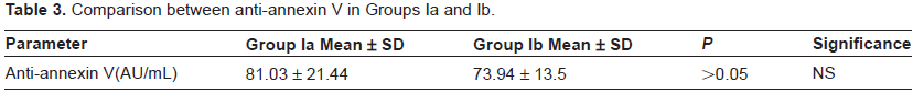
Anti-annexin V antibodies were present in 75% of patients (mean 83.46 ± 22.44 AU/mL) vs. 0% in the controls (mean 3.94 ± 4.5 AU/mL). Comparison between patients and controls as regards levels of anti-annexin V showed a highly significant difference (P < 0.001). However, comparison between disease subtypes (Groups Ia and Ib) showed no statistical significance (P > 0.05) (Tables 2 and 3).
Comparison between anti-annexin V in Groups I and II.

Comparison between anti-annexin V in Groups Ia and Ib.
Furthermore, the 20 SSc patients were re-classified into two groups according to the presence of severe ischemic manifestations including digital ulcers, gangrene and amputation.
Comparison between Group Is and Im as regards anti-annexin V antibodies.

NFC was performed for all patients and controls. It showed five patients with the early pattern (25%), eight (40%) with the active pattern and seven (35%) with the late pattern. As regards the data of the controls, all showed an organized pattern. No abnormal capillary loops, capillary haemorrhage or avascular areas were detected. Anti-annexin V antibodies were negative in the five patients with the early pattern and positive in all patients with the active and late patterns. On comparing titres of anti-annexin V between patients with active and late patterns, no statistical significance could be detected (P > 0.05) (Table 5).
Comparison of anti-annexin V between patients with active and late patterns.

For all patients and controls, Doppler echocardiography was done to assess pulmonary artery pressure and to evaluate pulmonary hypertension. In the patient group, the pulmonary artery pressure ranged from 28 to 45 mmHg with a mean ± SD of 35.00 ± 6.00 mmHg. Pulmonary hypertension was detected in five patients (25%). Pulmonary hypertension was not detected in any of the controls and their pulmonary artery pressure ranged between 20 and 30 mmHg with a mean value of 21.00 ± 5.00 mmHg. Comparing these results between patients and controls revealed a statistically significant difference (P < 0.05) (Table 6).
Comparison between Groups I and II as regards pulmonary artery pressure.

Coloured flow Doppler (CFD) of main renal, interlobar and cortical arteries was done for all patients and controls, and the RI was calculated. Comparison between the patients and controls as regards RI values of main renal, interlobar and cortical arteries showed a statistically significant difference (P < 0.05) (Table 7).
Comparison between patients and controls regarding the RI of the main renal, interlobar and cortical arteries.

Correlation studies between anti-annexin V and various parameters of the disease were performed. There was a significant positive correlation with each disease activity score (r = 0.53, P < 0.05), ACL (r = 0.79, P < 0.001) and RI of the main renal artery (r = 0.42, P < 0.05) (Table 8).
Correlation of anti-annexin V with variable disease parameters.

A receiver—operator characteristic curve (ROC) was drawn and analyzed to study the sensitivity and specificity of the anti-annexin V titre with its (positive and negative) predictive value and efficacy in discriminating patients with vascular damage from those without. At a cut-off value of 50 AU/mL, the anti-annexin V titre can discriminate cases with vascular damage from those without, with 62.5% specificity, 91.7% sensitivity, a negative predictive value (NPV) of 83.3%, a positive predictive value (PPV) of 78.6% and an efficacy of 80%.
Discussion
Systemic sclerosis is an autoimmune connective tissue disease associated with vascular damage, which can occur early in the course of the disease resulting in decreased capillary blood flow. Clinically overt symptoms of SSc include RP and fingertip ulcers. In addition, various internal organs can be involved, predominantly the lung, the kidneys and the heart. 10
This study was conducted on two main groups.
In this study, RP was the presenting symptom in 95% of patients included in the study. RP is known to be the presenting symptom in 95%–100% of SSc patients. Dysphagia and arthalgia are also common symptoms in SSc. Digital ulcers and pitting scars were detected in up to 67% of SSc patients and telangectasia was also frequently recorded.11, 12 ANA has been detected in 90% of SSc patients. AntiScl-70 and ACA are critical in differentiating both subtypes of SSc. ACL antibodies have been detected in SSc but with far less frequency than in other connective tissue disorders such as systemic lupus erythematosus and primary antiphospholipid syndrome. Recently, antifibrillin 1, anti-endothelial cells, anti-annexin V and anticollagen antibodies have been associated with SSc. 13
In this study, anti-annexin V antibodies were present in 75% of the patients (Group I) and was not detected in any of the controls (Group II) (mean 83.46 ± 22.44 vs. 3.94 ± 4.5 AU/mL). Comparison between both groups showed a highly significant statistical difference (P < 0.001). However, comparison of anti-annexin V antibodies in Groups Ia (diffuse) and Ib (limited) showed no statistical significance (P > 0.05).
To study the relationship of anti-annexin V antibodies with digital ischemia, patients were re-classified into two groups according to the presence of severe clinical manifestations of ischemia, including pitting scars, digital ulcers, gangrene and amputation.
Similar studies detected anti-annexin V antibodies in 18.2% of SSc patients and 75% of the patients showed ischemic manifestations in association with anti-annexin V antibodies. This could be explained by the fact that digital ischemia may not simply be explained due to thrombosis and that anti-annexin V antibodies could possibly be related to the pathogenesis of digital ischemia in SSc patients. 14
In addition, the anti-annexin V antibody titres in this study showed a highly significant positive correlation with ACL antibodies (r = 0.79, P < 0.001). Patients with antiphospholipid syndrome (APS) have been known to have a higher frequency of anti-annexin V antibodies, and thrombotic events have been reported more frequently in patients with positive anti-annexin V antibodies. 15 Furthermore, inhibition of annexin V binding to negatively charged phospholipids may be an additional pathogenic mechanism of APS. 16
The patterns of SSc was first described by NFC in 1981 as enlargement of the capillary loops, loss of capillaries, disruption of the orderly appearance of the normal capillary bed and capillary haemorrhages. 17 NFC parameters were found to correlate with the diffuse form of SSc, the degree of cutaneous involvement and the presence of AntiScl-70 antibodies. 18
Considering the SSc patterns obvserved in this study, 5% of patients had the early pattern, 75% had the active and 20% had the late pattern. Anti-annexin V antibodies were negative in the 5% of patients with the early pattern, and were positive in all patients with the active and late patterns. Comparison of anti-annexin V antibodies between patients with late and active patterns showed no statistical significance (P < 0.05); however, the mean levels of anti-annexin V antibodies in patients with the late pattern were somewhat higher.
In this study, Doppler echocardiography was done to assess pulmonary artery pressure and to evaluate pulmonary hypertension. In Group I, the pulmonary artery pressure ranged from 28 to 45 mmHg with a mean ± SD of 35.00 ± 6.00 mmHg. Pulmonary hypertension was detected in five patients (25%). Pulmonary hypertension was not detected in any of the controls. Comparison between patients and controls revealed a significant difference as regards pulmonary artery pressure (P < 0.05). However, correlation of anti-annexin V antibodies with mean pulmonary artery pressure in the patient group was of no statistical significance (P > 0.05).
Both types of lung disease, namely pulmonary hypertension and restrictive lung disease, have been detected in SSc. Vasculopathy causing pulmonary hypertension typically occurs in SSc.19, 20 Although the results of echocardiography are accurate and adequate for diagnosing advanced pulmonary hypertension, it cannot be relied upon to exclude pulmonary hypertension or to be used as a screening test for early cases. 21 On the other hand, screening for pulmonary hypertension by Doppler echocardiography enables early detection of pulmonary hypertension at a mild stage. 22
Considering the kidneys, comparison between the patients and controls as regards RI values of main renal, interlobar and cortical arteries showed a significant difference (P < 0.05). RI values tended to decrease from the main renal artery to the cortical artery.
In similar studies, RI values of renal vessels have been found to increase significantly in SSc patients compared to controls, and significant reduction of RI values between the interlobar and the cortical arteries of patients has been reported, which indicates an increased resistance to distal flow to the interlobar artery. 23 Renal vascular damage studied by CFD in SSc patients that are clinically free of nephropathy has detected latent and subclinical renal damage in these patients. 24 Other studies measuring the RI of renal and interlobar arteries in SSc patients with and without signs of renal involvement detected a significant increase in RI values in patients with signs of renal involvement as compared to those without. 25 However, in contrast to this study, a recent study on 44 patients with End stage renal failure indicated that circulating levels of anti-annexin V microparticles were increased compared with 32 healthy subjects, as were levels of microparticles derived from endothelial cells, platelets and erythrocytes. However, when arterial function was evaluated noninvasively in these patients, only endothelial microparticle levels correlated highly with loss of flow-mediated dilation and increased aortic pulse wave velocity. 26
In this study, all patients had apparently normal kidney functions, yet the significant difference between RI values of patients and controls suggests the presence of subclinical renal involvement in SSc patients, which could be due to a vasculitic process, and may progress rapidly to SSc renal crisis or renal failure. This puts more stress on the importance of early detection of renal vascular impairment in SSc. In this study, a significant positive correlation was detected between the level of anti-annexin V antibodies and RI of the main renal artery (r = 0.42, P < 0.05), further suggesting the association of anti-annexin V antibodies with renovascular damage in SSc and thus highlighting the value of its early detection in the follow-up of scleroderma patients.
In conclusion, SSc is associated with microvascular and macrovascular organ damage, especially in the pulmonary and renal arteries. Anti-annexin V antibodies are detected in SSc, with increased expression in patients with ischemic manifestations. Anti-annexin V antibodies correlate positively with the disease activity score, ACL antibodies and RI of the main renal artery, and could serve as a sensitive predictor of vascular damage in SSc.
Disclosures
This manuscript has been read and approved by all authors. This paper is unique and is not under consideration by any other publication and has not been published elsewhere. The authors and peer reviewers of this paper report no conflicts of interest. The authors confirm that they have permission to reproduce any copyrighted material.