
Abstract
Peripheral neutrophils are the predominant circulating leukocytes and an important component of innate and adaptive immune systems, which is a primary defense against cancer. MicroRNAs (miRNAs) can modulate neutrophil functions and play important roles in cancer pathogenesis by regulating neutrophil gene expression. To investigate if assessment of differential miRNA levels of peripheral neutrophils has the potential for diagnosis of non-small-cell lung cancer (NSCLC), we examine neutrophils of 15 patients with stage I NSCLC and 15 smokers without cancer. We identify five neutrophil miRNAs that have an abnormal level in patients with NSCLC versus smokers without cancer. In a training set of 82 patients with lung cancer and 73 controls, a set of two genes (miRs-26a-2-3p and 574-3p) are developed, producing 77.8% sensitivity and 78.1% specificity for NSCLC detection. Furthermore, in a testing set of 60 patients with lung cancer and 58 smokers, the performance of analyzing the two miRNAs for lung cancer detection is confirmed. This study for the first time shows that a neutrophil miRNA profile may serve as a new category of circulating biomarkers for the detection of NSCLC.
Introduction
The early diagnosis of lung cancer by low-dose computed tomography (LDCT) can significantly reduce the mortality. 1 However, >25% of smokers screened by LDCT have indeterminate pulmonary nodules (PNs), of which 96% are finally diagnosed to be false-positives. 1 Circulating biomarkers that can be used alone or complement CT for more sensitively and specifically detecting NSCLC are urgently needed.
Neutrophils are the predominant circulating leukocyte population, accounting for approximately 70% of peripheral leukocytes. 2 Neutrophils have emerged as an important component of effector and regulatory circuits in the innate and adaptive immune systems, 3 which are essential for immune reaction as a primary defense against cancer. Furthermore, the integration of immune and tumor cells promotes various molecular and cellular processes underlying multiple interrelated steps that define cancer initiation, development, and progression. 4 In addition, neutrophils contribute to the multiple steps of tumorigenesis by mediating cytotoxicity, tumor cell apoptosis, immunologically mediated tumor rejection, and antitumoral immune memory. 5 Previous studies have provided strong evidence for the existence of N1 (antitumoral) and N2 (protumoral) tumor-associated neutrophils (TANs).2,6–9 N1 neutrophils can kill tumor cells and thus play a beneficial and protective role for the host. Conversely, N2 neutrophils promote carcinogenesis in certain situations and are more closely associated with the invasive and metastatic effects in tumorigenesis. The assessment of TANs, particularly in surgically resected tumor tissue specimens, could predict disease-specific and overall survival in patients with metastatic and localized tumors. 2 However, there have been no reports regarding the use of peripheral neutrophils or the related molecular changes as potential circulating biomarkers for the detection of cancer at the early stage.
De novo induction of microRNAs (miRNAs) is part of the crucial regulatory circuits that control neutrophil gene expression 3 and hence modulates neutrophil functions.10–13 For instance, miR-223 can regulate innate immune genes whose functions are related to neutrophil biology. 14 Furthermore, the effect of miRNAs (miRs-491-3p, 34b, 595, 328, 1281, and 483-3p) has an important impact on neutrophil-related pathways. 15 miR-17 can regulate interleukin-8, a member of the cysteine-X-cysteine-chemokine, and has critical roles in neutrophil recruitment. 16 In addition, miRNAs (miRs-15b, 26a/b, 29a, 30b, 106b, and 93) govern the outcome of the neutrophil response to specific bacterial infections by mediating cytokine production. 15 miR-130a modulates transforming growth factor β1 control of neutrophil differentiation. 17 Moreover, both miR-17 and miR-31 can control neutrophil adherence on endothelial cells. 18 Therefore, the investigation of the neutrophil-related miRNAs could help understand neutrophil function and tumor biology. More importantly, charactering differential miRNA expression profiles of peripheral neutrophils might provide an immunomonitoring approach for tumorigenesis. Since neutrophils are the largest circulating leukocyte population in blood, hereby we investigate if miRNAs of peripheral neutrophils could be used as new circulation biomarkers for lung cancer.
Materials and Methods
Patient cohorts
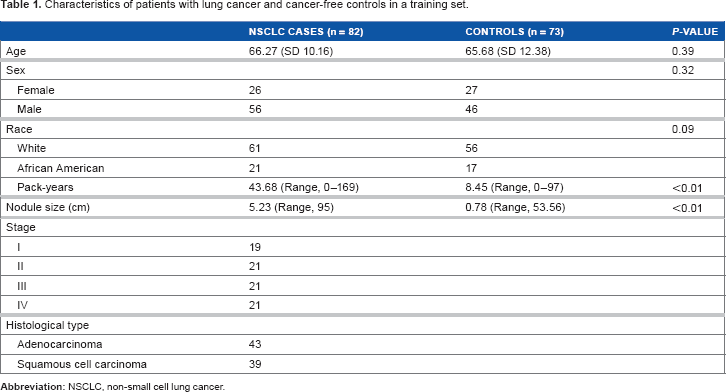
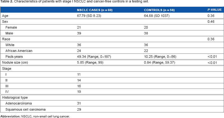
Our study was approved by the Institutional Review Boards of the Baltimore VA Medical Center and University of Maryland Medical Center. The research complied with the principles of the Declaration of Helsinki, and patients gave their written, informed consent to participate in this research. We recruited 157 patients with NSCLC (45 stage I, 35 II, 37 III, and 40 IV) and 146 smokers without any cancer, from whom peripheral blood was drawn as previously described. 19 The NSCLC cases consisted of two major types: squamous cell carcinoma and adenocarcinoma. These smokers without cancer had granulomatous inflammation (n = 72), nonspecific inflammatory changes (n = 53), or lung infections (n = 21). To delineate miRNA expression profiles in neutrophils of patients with lung cancer by an array platform, we used peripheral neutrophil specimens of 15 patients with stage I NSCLC and 15 smokers as an exploratory set. Furthermore, we randomly selected 82 patients with lung cancer with different stages and 73 smokers as a training set (Table 1). We used the training cohort to develop a panel of lung cancer biomarkers. We also selected 60 patients with stage I NSCLC and 58 cancer-free subjects as a testing set of cohort to confirm the performance of the biomarkers in the detection of NSCLC (Table 2).
Characteristics of patients with lung cancer and cancer-free controls in a training set.
Characteristics of patients with stage I NSCLC and cancer-free controls in a testing set.
Neutrophil isolation and processing
Peripheral neutrophils were isolated from blood as previously described.19–22 Briefly, a nucleated cell suspension was prepared using lymphocyte separation medium (MP Biomedicals, LLC). The cell pellet was lysed with ammonium chloride solution (Becton Dickinson and Company BD Biosciences). Neutrophils were isolated from the cells by using the Neutrophil Enrichment Kit (Stem Cell Technologies). The purity of the isolated cell populations was assessed by flow cytometry as previously described. 22 RNA was extracted from the cells as described in our earlier studies.19,23,24 Furthermore, we determined the purity, concentration, and integrity of RNA as described in our previously published articles.19,23,24 Briefly, the purity and concentration of RNA were determined by OD260/280 readings using a dual beam UV spectrophotometer (Eppendorf AG). A clean sample should have a 260/280 nm OD ratio of 1.8–2.0. RNA integrity was determined by capillary electrophoresis using the RNA 6000 Nano Lab-on-a-Chip kit and the Bioanalyzer 2100 (Agilent Technologies). A sample that had RNA integrity number of ≥7 was be considered as the one with very high quality. Only RNA extracts with RNA integrity number values >7 underwent in further analysis.
Realtime polymerase chain reaction-based microarray analysis of miRNAs
The analysis of miRNA expression profiles in neutrophils was performed by
Quantitative reverse transcriptase PCR
Quantitative reverse transcriptase (qRT)-PCR was carried out using a protocol that was developed in our laboratory.19,20 Briefly, 100 ng RNA was reversely transcribed by a T100 thermal cycler (Applied Biosystems) using miRCURY LNATM Universal cDNA Synthesis Kit (Exiqon). The thermocycler parameters were as follows: hold for 60 minutes at 42°C and for five minutes at 95°C. For qPCR analysis, after 40x dilution, 4 μL of cDNA was combined with 5 μL of miRCURY LNATM Universal RT miRNA PCR ExiLENT SYBR Green master mix (Exiqon) and 1 μL PCR primers set to produce a PCR reaction in a total volume of 10.0 μL. qPCR was carried out on an CFX96 thermocycler (Bio-Rad) at 95°C for 10 minutes, followed by 40 cycles of 95°C for 15 seconds and 60°C for one minute. qPCR data were analyzed by using the Manager software (Bio-Rad) with an automatic
Statistical analysis
We used Kruskal-Wallis test for comparing continuous variables or significance test for Spearman's rank correlation coefficient to determine the relations of the miRNAs and clinicopathologic and demographic characteristics of the subjects. We created receiver operating characteristic (ROC) curve and calculated the area under the ROC (AUC) value of each miRNA using numerical integration of the ROC curve. We utilized AUC to decide the accuracy of a biomarker on its capacity to differentiate cases from controls. We used Pearson's correlation analysis to evaluate correlation between the miRNAs.
Results
Identifying miRNAs that showed a differential expression level in peripheral neutrophils of lung cancer patients versus cancer-free subjects
Purity of the isolated peripheral neutrophils was greater than 95% as assessed by flow cytometry. Extracted RNA from the neutrophils was very pure as demonstrated by a 260/280 ratio of 1.8–2.0 and had very high quality as demonstrated by a RNA integrity number of ≥7. The measured miRNA expressions by using miRNA array in the replicates of each sample were highly correlated (all
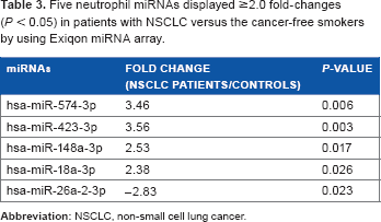
Five neutrophil miRNAs displayed ≥2.0 fold-changes (
Developing a panel of neutrophil miRNA biomarkers
Using qRT-PCR, we validated the results produced from the above miRNA array in a training set of 82 patients with lung cancer and 73 controls. All targeted five miRNAs showed ≤35
Expression levels of the miRNAs in neutrophils of patients with NSCLC versus cancer-free smokers by using qRT-PCR.
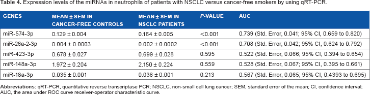
The two neutrophil genes displayed AUC values of 0.71–0.74 in differentiating patients with lung cancer from controls. Interestingly, the use of the two neutrophil miRNAs in combination created an AUC value of 0.81 (Fig. 2), being considerably higher than that of any single one used alone. There was no significant correlation between the two miRNAs’ level (
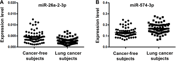
Expression levels of two miRNAs in neutrophil samples of 82 cancer-free smokers and 73 patients with NSCLC. The two miRNAs (A and B) have statistically significantly different levels in the patients with NSCLC versus cancer-free smokers (all
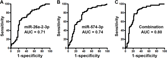
ROC curve analysis of expression levels of two miRNAs (miRs-26a-2-3p and -574-3p) in neutrophils of 82 patients diagnosed with NSCLC and 73 cancer-free individuals. miRs-26a-2-3p and -574-3p produce 0.71–0.74 AUC values (A and B), being significantly lower than 0.81 AUC created from the combined use of the two miRNAs (C) (
Validating the neutrophil miRNA biomarkers in a testing cohort
The use of the two neutrophil miRNAs in combination generated 78.3% sensitivity and 77.6% specificity in differentiating patients with all-stage lung cancer from cancer-free subjects (Supplementary Table 2). Combined use of the two genes created a higher sensitivity for the detection of advanced-stage (III–IV) NSCLC compared with stage I-II disease (82.9% vs 68.0% sensitivity,
Discussion
miRNAs have crucial functions in tumorigenesis. With the objective of developing circulating cancer biomarkers, abundant studies have been done by detecting cell-free miRNAs that are released from tumor cells in blood.19,20,25–30 Though displaying promise, the analysis of the extracellular genes in serum or plasma has some difficulties, limiting its use in the clinical practice. These mainly consist of 1) low recovery of miRNAs in plasma or serum, 31 2) the discharge of miRNAs in plasma or serum by hemolysis of RBCs, creating nonspecific consequences, and 3) sources of inconsistency of the cell-free miRNAs may cause varying or even opposing results for the identification of the same type of cancer. 32 The difficulty turn out to be more complex by the fact that in blood, miRNAs are either associated with proteins, such as argonaute32,33 and lipoproteins, 34 or enclosed within cellular fragments designated as exosomes, microparticles, microvesicles, or extracellular vesicles.35,36
The analysis and development of circuiting neutrophil miRNAs as blood-based biomarkers may address the above challenges in the development of the cell-free miRNAs because it is a cell-based approach. Furthermore, peripheral neutrophils make up 70% of white blood cells and thus can produce large amounts of RNA for reliable analysis of miRNAs. In addition, our results show that extracted RNA from the neutrophils has very high quality. Moreover, purity of neutrophils was greater than 95%, from which RNA is specifically isolated and may not contain RBC miRNAs and other blood components. Therefore, neutrophil miRNA profile may serve as a new category of circulating biomarkers for the detection of NSCLC.
Of the two miRNAs, only miR-26a-2-3p was previously found to be associated with lung cancer and had an elevated expression level in lung tumor tissues.19,37–39 However, our present study showed that miR-26a-2-3p exhibited a low level in neutrophils of patients with NSCLC compared with cancer-free controls. The contradictory observations suggest that the miRNA expression change in neutrophils may not simply be a reflection of that in the lung cancer tissues or mimic that in the primary tumors. Furthermore, previous reports including our own have found that numerous cell-free miRNAs released from primary cancer sites have the potential for lung cancer diagnosis. 20 Yet, no overlap is observed between the neutrophil miRNAs of lung cancer versus the plasma or serum miRNAs from lung cancers.20,40 The neutrophil miRNAs may not be influenced by circulating cancer cells. Therefore, the assessment of the neutrophil miRNAs could present a different strategy for lung cancer diagnosis. In addition, differing from the analysis of circulating extracellular molecules that are dependent on substantial cancer burden, the determination of neutrophil miRNA expressions may function as a better surrogate window into cancer status, as the dysregulation of neutrophil miRNAs is involved in every step of tumorigenesis, such as cytotoxicity, tumor cell apoptosis, immunologically mediated tumor rejection, and antitumoral immune memory.3,10–13 Nevertheless, carrying out a new study to compare miRNAs of matched neutrophils, plasma, and cancer tissues of the same patients is necessary to investigate if the neutrophil miRNAs are independent from those in blood and cancers of the patients. Furthermore, analyzing the potential neutrophil miRNA biomarkers in different types of malignancy to determine if they are specific to lung cancer is also required.
Neutrophils play a dual role in tumorigenesis through innate and adaptive immune systems that can recognize and remove malignant cells, a process called
miR-26a family has been suggested to play an important function in modulating neutrophil biology. For example, miR-26a was one of the abundant miRNAs in neutrophils.41,42 miR-26a expression could be preferentially induced by
The study has some limitations. First, we use an array that only analyzes 372 miRNA genes to delineate the neutrophil miRNAs whose changes are associated with lung cancer. Only 141 miRNAs could be detectable in the neutrophil specimens. In addition, from the 141 genes, only two are finally found to have the potential as biomarkers. The two miRNAs used together has 77.78% sensitivity and 78.08% specificity for the detection of all-stage NSCLC. However, the efficiency of the biomarkers is not enough to be used in laboratory settings. Particularly, the diagnostic performance of the two biomarkers for the early-stage NSCLC is not sufficient because the sensitivity for stage I–II NSCLC is only 68.0%. To address the concern, we are using whole-genome next-generation sequencing to identify new neutrophil miRNAs of lung cancer that can be added in the assay for precisely diagnosing lung cancer. Second, the neutrophil samples are acquired from the hospital-based patients with clinical diagnosis. Future validation of the biomarkers in a prospective and multisite LDCT lung cancer screening trial is needed.
Conclusion
In conclusion, we find a differential miRNA expression profile in peripheral neutrophils of patients with lung cancer. Furthermore, two circulating neutrophil miRNA biomarkers are developed that have the potential to be used for the detection of lung cancer. Nevertheless, finding additional neutrophil miRNA biomarkers that can be added to the two biomarkers and incorporating other types of biomarkers with the neutrophil miRNA biomarkers for lung cancer early detection are required. Moreover, given the contribution of neutrophil miRNAs to both the initiation and the progression of tumors, we believe that different miRNA profiles of neutrophils could be also used for the development of new circulating biomarkers in other cancer diseases.
Author Contributions
Conducted the experiments and participated in data acquisition and interpretation: JM, NL, YL, and CG. Conducted the study design, coordination, and prepared the manuscript: FJ. All authors read and approved the final manuscript.
Supplementary Materials
Footnotes
Acknowledgments
The authors thank the Biostatistics Shared Service of University of Maryland School of Medicine for statistically analyzing the data.