
Abstract
Capillary hemangioma is a common benign tumor which can occur everywhere in the whole body, however its occurrence in posterior mediastinum is extremely rare, and to the best of our knowledge less than 20 cases have been reported in the English literature so far. Here in we report a 65-year-old lady who presented with prolonged cough and diagnosed to have a posterior mediastinal mass. Before operation, according to the site of tumor, it has been diagnosed as neurofibroma. It is very important to consider hemangioma before operation to reduce surgical complications, and it should be in the differential diagnosis of posterior mediastinal masses.
Keywords
Introduction
Mediastinal hemangioma is a rare lesion, and less than 0.5% of the mediastinal masses are hemangioma. 1 Most of these reported mediastinal hemangiomas have been in anterior part and of cavernous type. 2
Herein we report an extremely rare case of posterior mediastinal mass that has been operated with the primary impression of neurofibroma and was diagnosed as capillary hemangioma after pathologic examination of the resected mass.
Case Report
A 65-year old female, case of diabetes mellitus and hypertension, referred with chief complaint of productive cough for one year.
Physical examination and routine blood tests were unremarkable. No positive previous history has been noted, i.e. there has been no history of surgery or previous mass or tumor.
Plain chest-X-ray radiography showed a well defined round opacity in right side of mediastinum (Figure 1A). Spiral chest computed tomography with contrast showed a 5×5 cm mass in close contact to posterior chest wall and vertebra with peripheral enhancement and mild pleural effusion. Pressure effect on right main bronchus and mild displacement of esophagus was also present (Figure 1B).
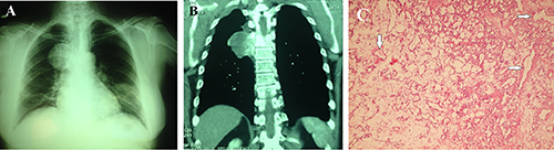
A) Chest X-ray shows a mediastinal mass. B) Computed tomography scan shows a large mass lesion measuring 5×5 cm in posterior segment of mediastinum. C) Sections from posterior mediastinal mass show vascular channels lined by normal endothelial cells (Hematoxylin & Eosin, 250×).
Tru-cut biopsy was unsuccessful and no adequate tissue was yielded for definite diagnosis. It showed some mature adipose tissue and a few spindle-shaped cells and vessels. With the preliminary diagnosis of neurogenic tumor (neurofibroma) thoracotomy and complete excision was done for the patient and the mass sent for pathology.
Histopathology revealed that the mass consisted of small vessels of capillary caliber in vague lobular configuration with no atypia and low mitotic figures. There has been no hemorrhage and thrombosis (Figure 1C).
Diagnosis of capillary hemangioma was made. Now after 6 months, she is doing well and completely frees of symptoms.
Discussion
Mediastinal masses span a wide histopathological spectrum. They are divided into anterior, middle and posterior compartments. Anterior mediastinal tumors account for 50% of all mediastinal masses, including thymoma, teratoma, thyroid lesions and lymphoma. Masses of the middle mediastinum are typically congenital cysts while those arising in the posterior mediastinum are often neurogenic tumors. 3
The first case of mediastinal hemangioma has been reported in 1914 by Shannon. 4 After that, more than 100 cases of mediastinal hemangioma have been reported in the English literature. 5
Almost all of the previously reported cases have been cavernous type in anterior mediastinum and to the best of our knowledge less than 20 cases of posterior mediastinal hemangioma have been reported in the English literature so far. Between these posterior mediastinal hemangiomas only 6 cases have been capillary type hemangioma.1,3,4
Most of the reported posterior mediastinal hemangiomas have been in female patients, in adults, but rare cases of pediatric patients have been reported. 6 Our patient has been a 65-year old lady who presented with prolonged cough, also, the most common presenting symptom in the previous cases has been pulmonary symptoms such as cough, and chest pain. 7
The most challenging point in posterior mediastinal capillary hemangioma has been preoperative diagnosis, none of the previous cases had preliminary diagnosis of hemangioma and almost all of the cases have been operated with the preoperative diagnosis of neurogenic tumor such as neurofibroma.8–10
Calcification, and phlebolits are diagnostic signs in mediastinal hemangiomas, however these findings have been reported in less than 20% of the cases. 10
Radiologically mediastinal hemangioma shows homogenous signal intensity which is very similar to neurogenic tumors such as paraganglioma and pheochromocytoma. 10 In MRI (magnetic resonance imaging) differences in signal intensity on T-1 and T-2 weighted images can be helpful and also markedly high intensity on fat suppression T-2 weighted image might be a characteristic finding. 5 Our patient has been operated with preliminary diagnosis of neurofibroma, mostly because of posterior mediastinal location of the mass.
Conclusions
The purpose of surgery in most of previous cases has been both diagnosis and treatment. 8 Endovascular embolization is also another option for removing mediastinal hemangioma, which can also reduce blood loss during surgery, 7 however, it is often difficult to diagnose mediastinal hemangioma before surgery. 10
Histologically, most of the capillary hemangiomas in the mediastinum have been lobulated, separated by thin fibrovascular septae. The tumor cells have been bland looking with no atypia, and low or no mitosis. 11
Prognosis of capillary hemangioma after resection is excellent. There has been no recurrence in long follow up. 12
Table 1 shows the characteristics of posterior mediastinal hemangioma reported so far.
Characteristics of posterior mediastinal hemangioma reported so far.

y, years; m, months; w, weeks; M, male; F, female; CHD, coronary heart disease; TOF, tetralogy of Fallot.
As a conclusion, hemangioma and vascular tumors can occur anywhere in the body, so it should also be in the differential diagnosis of posterior mediastinal masses, because it can cause surgical complications such as bleeding during operation.